Systems and Methods for Robust Learning Based Annotation of Medical Radiographs

a learning-based, medical radiograph technology, applied in the field of anatomy detection, can solve the problems of inaccurate recognition, affecting the accuracy of feature-based recognition techniques, and affecting the accuracy of medical radiographs, and achieve the effect of low accuracy

Active Publication Date: 2010-11-11
SIEMENS HEALTHCARE GMBH
View PDF6 Cites 81 Cited by
  • Summary
  • Abstract
  • Description
  • Claims
  • Application Information

AI Technical Summary

Benefits of technology

[0026]A computer system includes a processor and a program storage device readable by the computer system, embodying a program of instructions executable by the processor to perform method steps for locating a region of interest. The method includes acquiring a medical image; automatically detecting a set of local feature candidates representing potential anatomical landmarks from the medical image; assessing the accuracy of each local feature candidate using multiple combinations of the other local feature candidates and removing a local feature candidate that is assessed to have the lowest accuracy; repeating the assessing and removing step until only a predetermined number of local feature candidates remain; locating a provided region of interest (ROI) from within the medical image based on the remaining predetermined number of local feature candidates.

Problems solved by technology

Recognizing anatomical structures within digitized medical images presents multiple challenges.
However, feature based recognition techniques can suffer from accuracy problems.
While CT imaging is relatively safe, it does involve exposure to ionizing radiation, which could become harmful in patients.
However, the more precise the field is, the faster the scan can be performed and the less the subject is exposed to potentially harmful ionizing radiation.
However, this manual determination may take an amount of time that is noticeable to the subject, and as such, there is a greater possibility that the subject may shift position between the acquisition of the topogram and the acquisition of the CT scan within the manually determined scanning field.
Moreover, the manually selected scanning field may be slightly different each time a CT scan is performed and thus multiple CT scans, such as follow-up studies of the same patient and / or cross-patient comparisons, may be more difficult to compare owing to the inherent inconsistency of the manual field selection.
In addition to the above-described difficulties in correctly identifying a scanning field for a medical image study, annotation of the ever-increasing number of medical images is an overwhelming task.
Manually annotating these images is costly and error-prone, which means that automatic annotation algorithms are needed and must to be able to perform the task reliably and efficiently.
This is particularly true for radiograph images, although similar issues exist for other imaging modalities.
A great challenge for automatic medical image annotation is the large visual variability across patients in medical images from the same anatomy category.
In some cases, diseases or artifacts can render an anatomy unrecognizable even by human eyes.

Method used


Image

  • Systems and Methods for Robust Learning Based Annotation of Medical Radiographs
  • Systems and Methods for Robust Learning Based Annotation of Medical Radiographs
  • Systems and Methods for Robust Learning Based Annotation of Medical Radiographs

Examples


Embodiment Construction

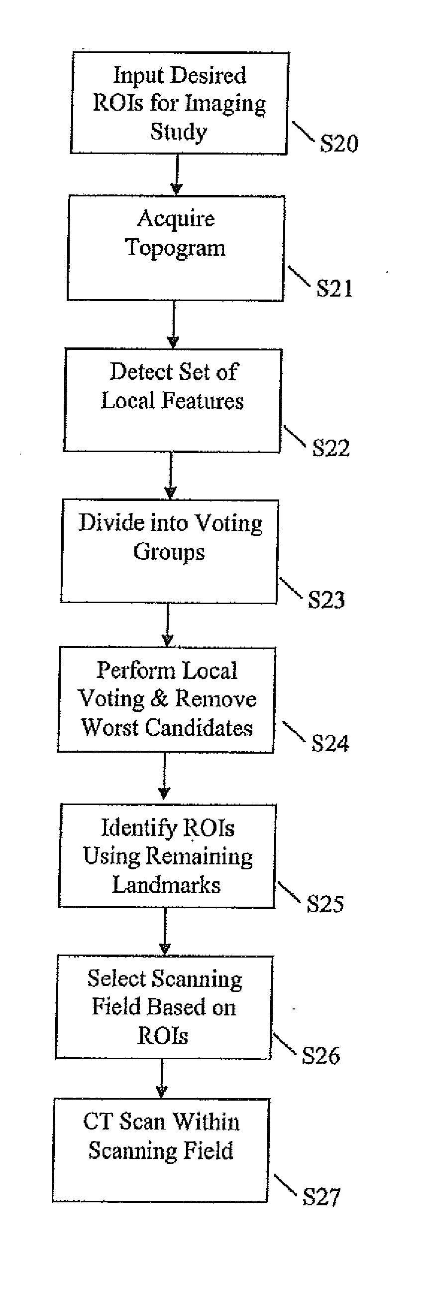
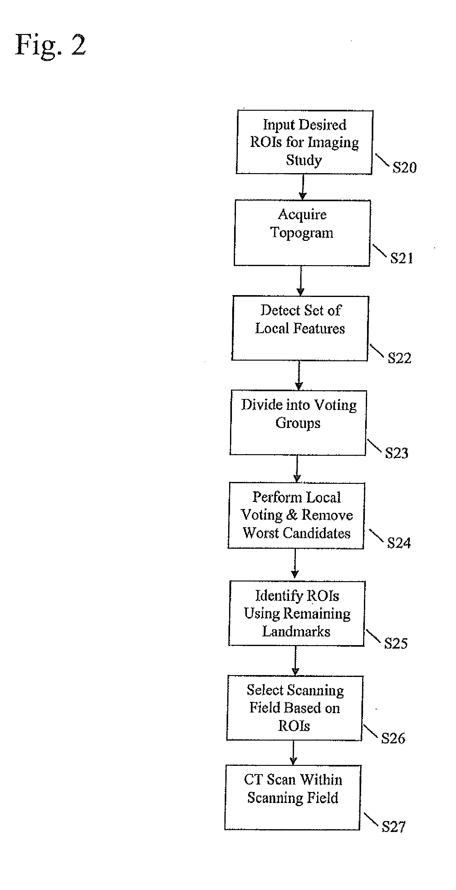
[0036]In the following description, numerous specific details are set forth such as examples of specific components, devices, methods, etc., in order to provide a thorough understanding of embodiments of the present invention. It will be apparent, however, to one skilled in the art that these specific details need not be employed to practice embodiments of the present invention. In other instances, well-known materials or methods have not been described in detail in order to avoid unnecessarily obscuring embodiments of the present invention. While the invention is susceptible to various modifications and alternative forms, specific embodiments thereof are shown by way of example in the drawings and will herein be described in detail. It should be understood, however, that there is no intent to limit the invention to the particular forms disclosed, but on the contrary, the invention is to cover all modifications, equivalents, and alternatives falling within the spirit and scope of th...


PUM


Abstract

Systems and methods for performing a medical imaging study include acquiring a preliminary scan. A set of local feature candidates is automatically detected from the preliminary scan. The accuracy of each local feature candidate is assessed using multiple combinations of the other local feature candidates and removing a local feature candidate that is assessed to have the lowest accuracy. The assessing and removing steps are repeated until only a predetermined number of local feature candidates remain. A region of interest (ROI) is located from within the preliminary scan based on the remaining predetermined number of local feature candidates. A medical imaging study is performed based on the location of the ROI within the preliminary scan.

Description

CROSS-REFERENCE TO RELATED APPLICATION[0001]The present application is a Continuation-hi-Part Application of U.S. patent application Ser. No. 12 / 334,898, filed Dec. 15, 2008, which claims the benefit of U.S. Provisional Application No. 61 / 016,313, filed Dec. 21, 2007, which applications are hereby incorporated herein by reference in their entirety. The present application additionally claims the benefit of U.S. Provisional Patent Application No. 61 / 181,035, filed May 26, 2009, which application is hereby incorporated herein by reference in its entirety.TECHNICAL FIELD[0002]The present disclosure relates to anatomy detection and, more specifically, to robust anatomy detection though local voting and prediction.BACKGROUND[0003]The field of medical imaging has seen significant advances since the time X-Rays were first used to determine anatomical abnormalities. Medical imaging hardware has progressed in the form of newer machines such as Medical Resonance Imaging (MRI) scanners, Comput...

Claims


Application Information

Patent Timeline
Patent Type & Authority Applications(United States)
IPC IPC(8): G06K9/46G06K9/00G06V10/25G06V10/764G06V10/771
CPCA61B6/032A61B6/469A61B6/488G06K9/3233G06K9/6231G06K9/6267G06T2207/30048G06T7/0042G06T7/0083G06T2207/10081G06T2207/20104G06T2207/20164G06K2209/05G06T7/73G06T7/12G06V10/25G06V2201/03G06V10/771G06V10/764G06F18/2115G06F18/24
Inventor PENG, ZHIGANGTAO, YIMOZHOU, XIANG SEANZHAN, YIQIANGKRISHNAN, ARUN
Owner SIEMENS HEALTHCARE GMBH
Why Eureka
Try Eureka