Mammography method and mammography apparatus

a mammography and method technology, applied in the field of mammography method and mammography apparatus, can solve the problems of limiting the use of agents, affecting the ability to detect such tumors, and affecting the ability to detect tumors

Inactive Publication Date: 2010-11-18
SIEMENS HEALTHCARE GMBH
View PDF3 Cites 49 Cited by
  • Summary
  • Abstract
  • Description
  • Claims
  • Application Information

AI Technical Summary

Benefits of technology

[0009]An object of the invention is to provide a mammography method in which the risks explained above accompanying the x-ray examinations implemented with the administration of a contrast agent are reduced. A further object of the invention is based to provide a mammography apparatus suitable to implement such a mammography method.

Problems solved by technology

Since the tissue slices situated one after another in the direction of the x-ray beam are superimposed in such a projection, strongly absorbent benign structures can overlay a malignant tumor and hinder the ability to detect such a tumor.
The contrast agents that are used are normally toxic and can be linked to unwanted side effects, such that their use should be limited to a minimum.

Method used


Image

  • Mammography method and mammography apparatus
  • Mammography method and mammography apparatus
  • Mammography method and mammography apparatus

Examples


Embodiment Construction

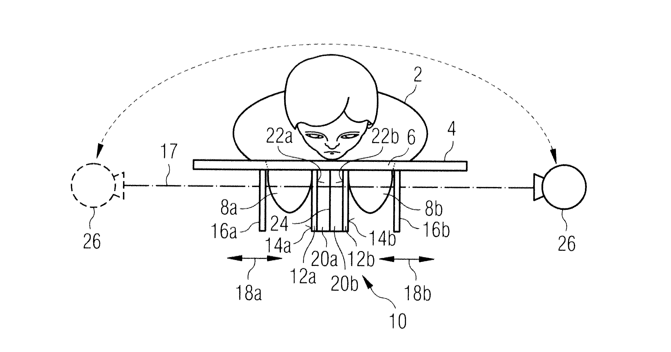
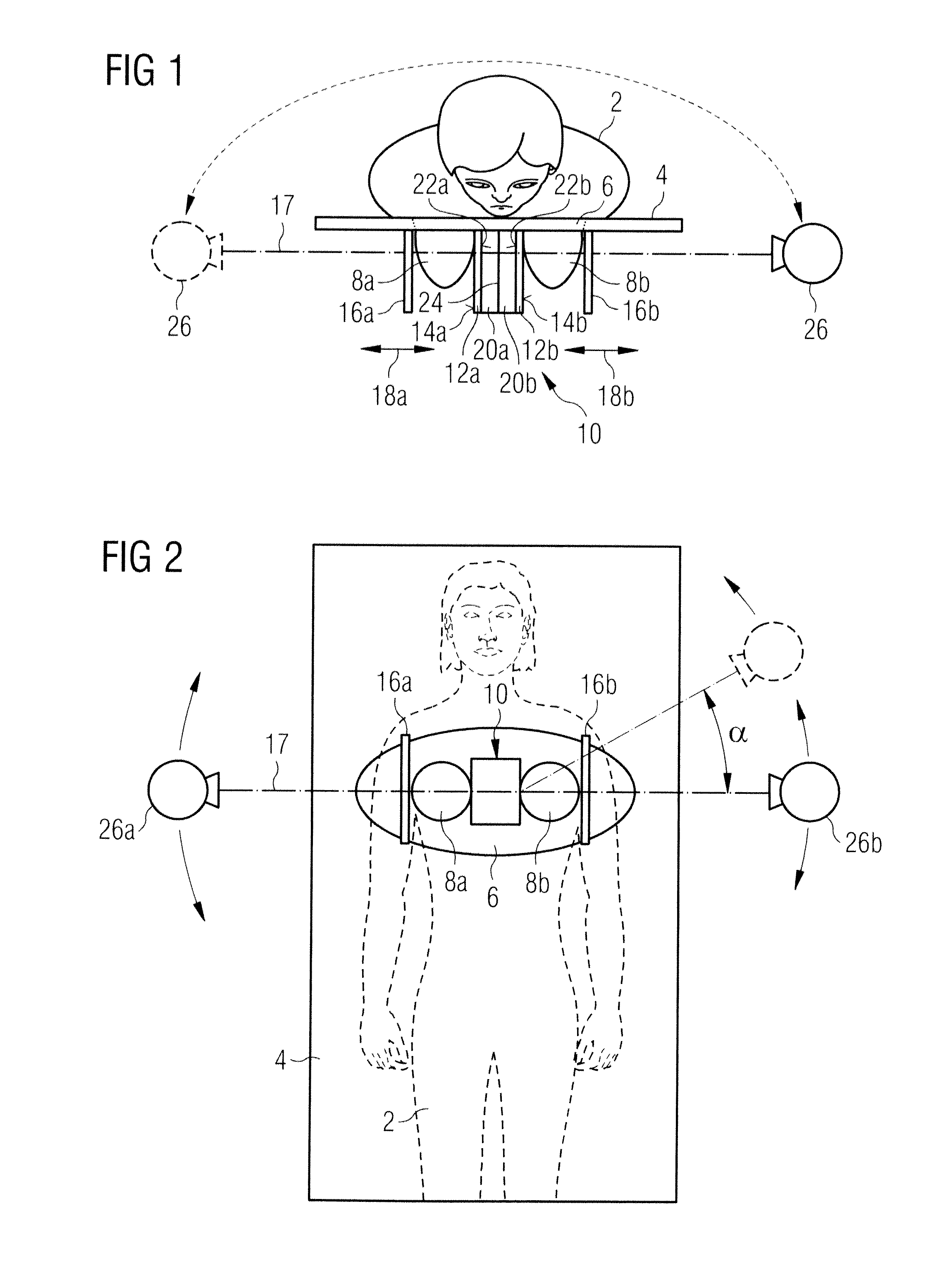
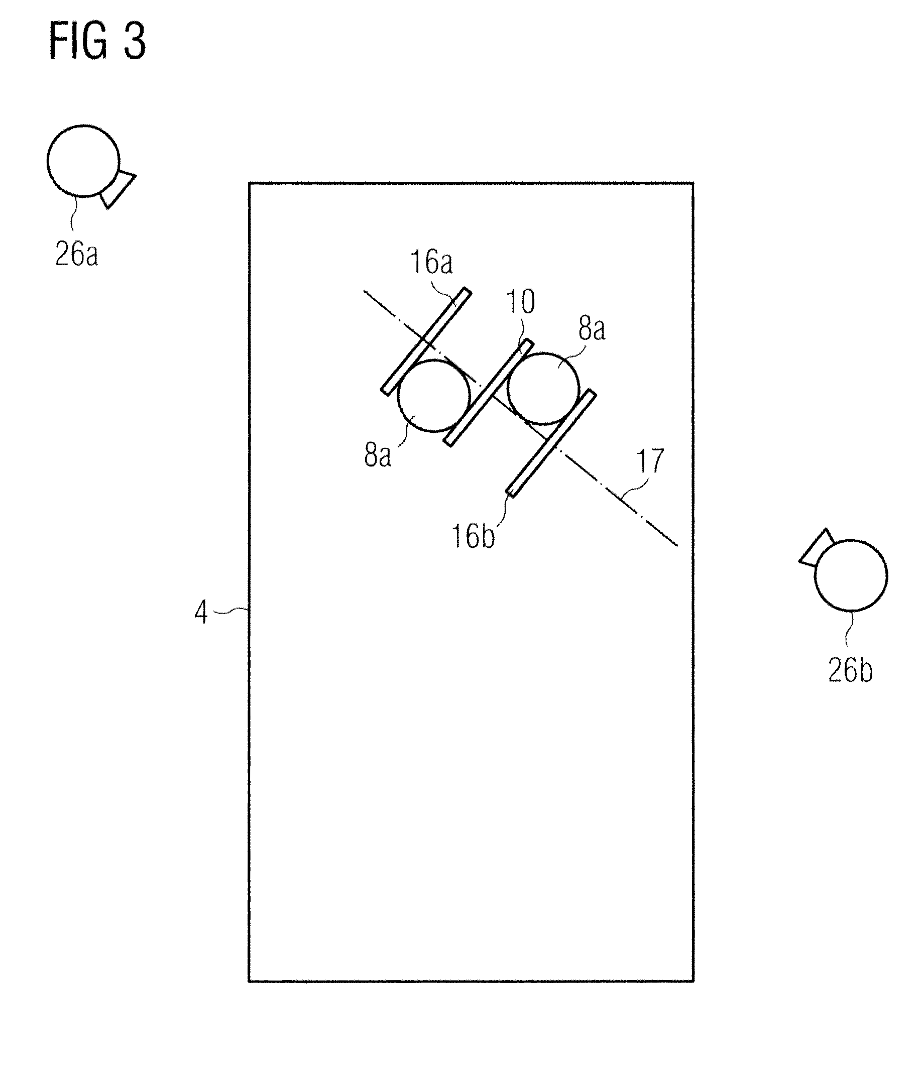
[0018]According to FIG. 1, an examination subject 2 is located in ventral position on a patient table 4. In the patient table 4 is an opening through which both breasts 8a, b of the examination subject 2 protrude so that they project beyond the underside of the patient table 4. A support unit 10 that possesses two support plates 12a, 12b situated opposite one another is arranged below the patient bearing table 4, approximately central relative to the opening 6. The bearing plates 12a, 12b form support surfaces 14a and 14b situated opposite one another and respectively facing toward respective compression plates 16a, 16b. Compression plates 16a and 16b are aligned with their flat sides parallel to one another and are supported such that they can be displaced perpendicularly to their flat sides in the direction of a compression axis 17 (horizontal in the example). When the patient 2 lies face down on the table 4, the breast 8a is between the compression plate 16a and the support surfa...


PUM


Abstract

In a mammography method and a mammography apparatus to generate x-ray images of a breast, a support unit is positioned between the breasts. In the support unit two x-ray detectors are arranged with acquisition surfaces that are flat and parallel to one another. The acquisition surfaces face away from one another and respectively toward one of the breasts. At least one x-ray image of each breast is acquired while both breasts are simultaneously pressed against the support unit.

Description

BACKGROUND OF THE INVENTION[0001]1. Field of the Invention[0002]The present invention concerns a mammography method and a mammography apparatus suitable to implement the method.[0003]2. Description of the Prior Art[0004]Mammography is an x-ray examination of the breast with the goal of detecting tumors at an earliest possible stage. Through continuous improvement of mammography techniques it is sought to generate x-ray images with high significance in order to differentiate benign tissue variations from malignant variations and to reduce the number of incorrect findings, i.e. the number of suspicious findings that are caused by non-malignant variations and the number of undetected malignant tumors. In conventional x-ray mammography, a single two-dimensional image of the compressed breast is generated at a single projection direction. Since the tissue slices situated one after another in the direction of the x-ray beam are superimposed in such a projection, strongly absorbent benign ...

Claims


Application Information

Patent Timeline
Patent Type & Authority Applications(United States)
IPC IPC(8): A61B6/04
CPCA61B6/025A61B6/0435A61B6/4007A61B6/502A61B6/466A61B6/482A61B6/4241
Inventor ELIASSON, EVA
Owner SIEMENS HEALTHCARE GMBH
Why Eureka
Try Eureka