Method and System for Database-Guided Lesion Detection and Assessment

Inactive Publication Date: 2011-01-13
SIEMENS AG
View PDF12 Cites 34 Cited by
  • Summary
  • Abstract
  • Description
  • Claims
  • Application Information

AI Technical Summary

Problems solved by technology

However, conventional clinical practice exhibits a number of limitations.
The restriction to only a subset of target lesions in mainly due to the fact that manual assessment and size measurement o

Method used


Image

  • Method and System for Database-Guided Lesion Detection and Assessment
  • Method and System for Database-Guided Lesion Detection and Assessment
  • Method and System for Database-Guided Lesion Detection and Assessment

Examples


Embodiment Construction

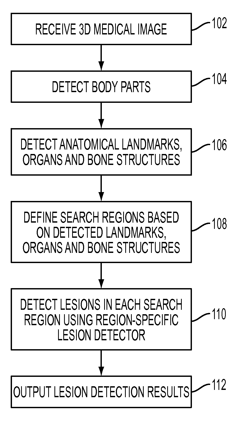
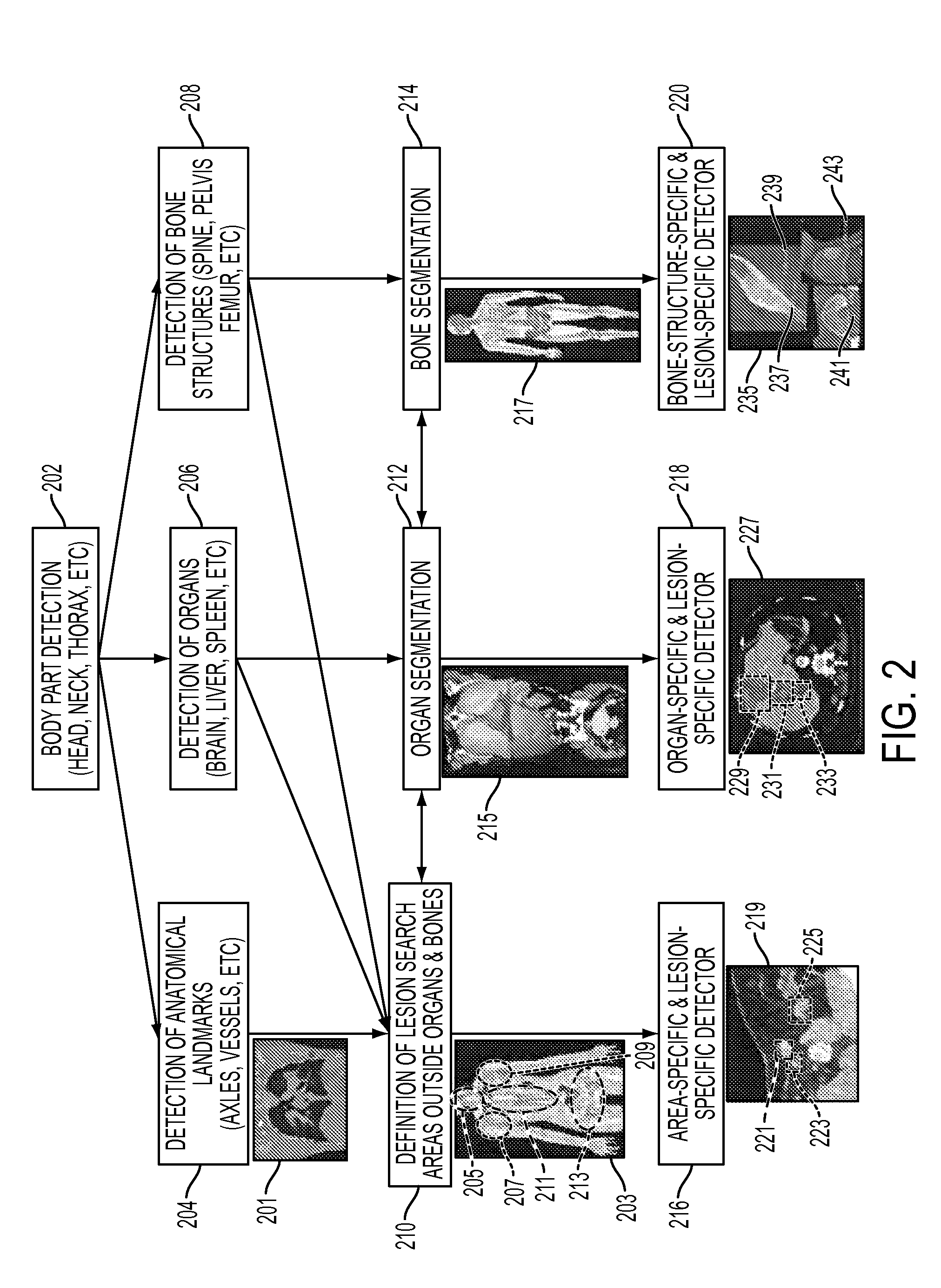
[0020]The present invention is directed to a method and system for automatic detection of lesions in 3D medical images, such as computed tomography (CT) and magnetic resonance (MR) images. A digital image is often composed of digital representations of one or more objects (or shapes). The digital representation of an object is often described herein in terms of identifying and manipulating the objects. Such manipulations are virtual manipulations accomplished in the memory or other circuitry / hardware of a computer system. Accordingly, it is to be understood that embodiments of the present invention may be performed within a computer system using data stored within the computer system.

[0021]Embodiments of the present invention provide methods for lesion detection and assessment in 3D medical image data, such as CT and MR data. The automatic lesion detection method described herein can be used to detect lesions in various parts of the body including, but not limited to, lymph nodes, o...


PUM


Abstract

A method and system for automatically detecting lesions in a 3D medical image, such as a CT image or an MR image, is disclosed. Body parts are detected in the 3D medical image. Anatomical landmarks, organs, and bone structures are detected in the 3D medical image based on the detected body parts. Search regions are defined in the 3D medical image based on the detected anatomical landmarks, organs, and bone structures. Lesions are detected in each search region using a trained region-specific lesion detector.

Description

[0001]This application claims the benefit of U.S. Provisional Application No. 61 / 224,488, filed Jul. 7, 2009, the disclosure of which is herein incorporated by reference.BACKGROUND OF THE INVENTION[0002]The present invention relates to lesion detection in 3D medical images, and more particularly, to automatic database-guided lesion detection in medical images, such as computed tomography (CT) and magnetic resonance (MR) images.[0003]Tumor staging and follow-up examinations account for a large portion of routine work in radiology. Cancer patients are typically subjected to examinations using medical imaging, such as CT, MR, or positron emission tomography (PET) / CT imaging, in regular intervals of several weeks or months in order to monitor patient status or assess responses to ongoing therapy. In such examinations, a radiologist typically checks whether tumors have changed in size, position, or form, and whether there are new lesions. However, conventional clinical practice exhibits ...

Claims


Application Information

Patent Timeline
IPC IPC(8): G06K9/00
CPCG06K9/00362G06T7/0012G06T2207/30096G06T2207/20076G06T2207/20101G06T2207/10072G06V40/10
Inventor SUEHLING, MICHAELSOZA, GRZEGORZ
Owner SIEMENS AG
Why Eureka
Try Eureka