Medical training method and apparatus

a training method and medical technology, applied in the field of medical training methods and equipment, can solve problems such as the inability to reduce accuracy

Active Publication Date: 2011-07-14
INVENTIVE MEDICAL
View PDF15 Cites 47 Cited by
  • Summary
  • Abstract
  • Description
  • Claims
  • Application Information

AI Technical Summary

Problems solved by technology

Although TTE is considered a highly accurate procedure, the accuracy can be reduced because of obesity, chronic obstructive pulmonary disease, or chest-wall deformities.

Method used


Image

  • Medical training method and apparatus
  • Medical training method and apparatus
  • Medical training method and apparatus

Examples


Embodiment Construction

[0100]The various embodiments mentioned above will be described in further detail with reference to the attached figures.

[0101]First, conventional medical imaging devices will briefly be described with reference to FIGS. 1 to 3.

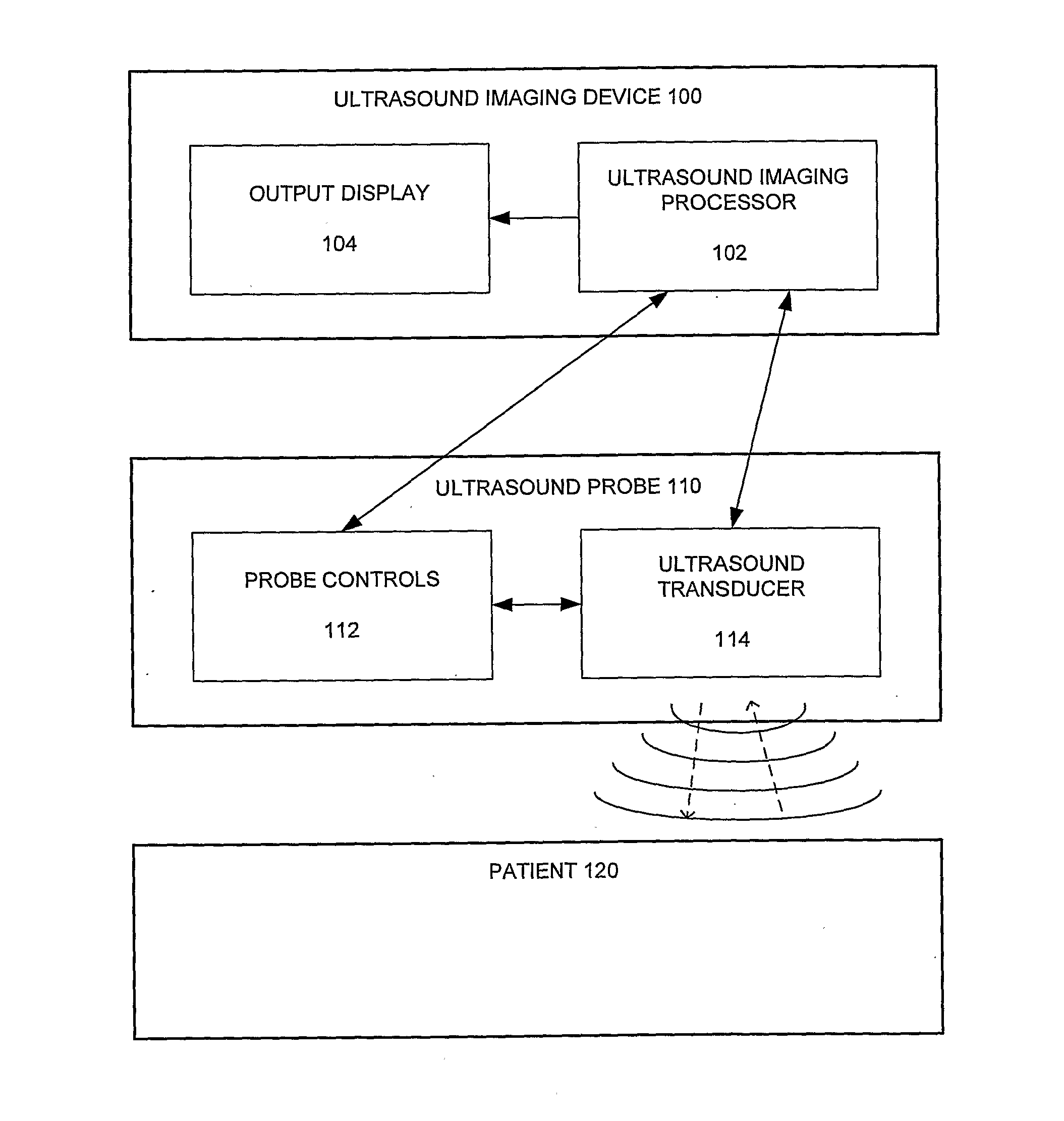
[0102]FIG. 1 is an illustration of the operation of a conventional ultrasound scanning device.

[0103]An ultrasound imaging device 100 and an ultrasound probe 110 are used to image anatomical structures within the patient 120. The imaging device 100 includes an ultrasound imaging processor 102 for controlling the generation of appropriate ultrasound signals and for interpreting the received ultrasound reflections, and an output display 104 for outputting the result of the processing by the processor 102. The probe 110 may include probe controls 112 (as is discussed in more detail below), and an ultrasound transducer 114 for generating and receiving the ultrasound waves.

[0104]In use, input devices (not shown) can allow various properties of the ultrasound scan t...


PUM


Abstract

There is disclosed a method of simulating the output of a medical imaging device, the medical imaging device being operable to image an anatomical structure, and the method including: accessing model data representing a model of the anatomical structure; accessing selection data representing a selected region of the anatomical structure to be imaged; and processing the selection data and the model data to generate output data representing a simulation of the output of the medical imaging device when imaging the selected region.

Description

FIELD OF THE INVENTION[0001]The present invention relates generally to facilitating medical training, but has further applications.BACKGROUND OF THE INVENTION[0002]Medical imaging devices provide an important diagnostic tool for many medical applications. One example of a medical imaging device is an ultrasound scanner. Ultrasound scans are carried out by transmitting high frequency ultrasonic sound into a human body and received and processing reflections of the transmitted sound in order to create an image of an organ or other structure in the body.[0003]For diagnostic purposes, a medical professional performs an ultrasound scan using a hand-held probe (or more commonly referred to as a transducer) that is placed directly on and moved over the surface of the body. Essentially, the transducer transmits sound waves into the body and receives echoing waves that reflect from internal organs, fluids and tissues. The reflected waves are converted to corresponding electrical pulses that ...

Claims


Application Information

Patent Timeline
Patent Type & Authority Applications(United States)
IPC IPC(8): G06K9/00G06F17/50
CPCG09B23/285A61B8/44A61B8/58
Inventor MARTIN, COLIN BRUCEWRIGHT, SUSANSMITH, ANDREWCUBITT, ADAM WILLIAM JON
Owner INVENTIVE MEDICAL
Why Eureka
Try Eureka