Diagnostic imaging apparatus, diagnostic ultrasonic apparatus, and medical image displaying apparatus

a diagnostic ultrasonic and imaging apparatus technology, applied in tomography, applications, instruments, etc., can solve the problems of reducing the display area of ultrasonic images and not always using the display area effectively during examination

Active Publication Date: 2011-11-17
TOSHIBA MEDICAL SYST CORP
View PDF3 Cites 16 Cited by
  • Summary
  • Abstract
  • Description
  • Claims
  • Application Information

AI Technical Summary

Problems solved by technology

With such a conventional technology, however, if graphics such as switches, icons, and tabs are constantly displayed on the monitor, these graphics reduce the display area of an

Method used


Image

  • Diagnostic imaging apparatus, diagnostic ultrasonic apparatus, and medical image displaying apparatus
  • Diagnostic imaging apparatus, diagnostic ultrasonic apparatus, and medical image displaying apparatus
  • Diagnostic imaging apparatus, diagnostic ultrasonic apparatus, and medical image displaying apparatus

Examples


second embodiment

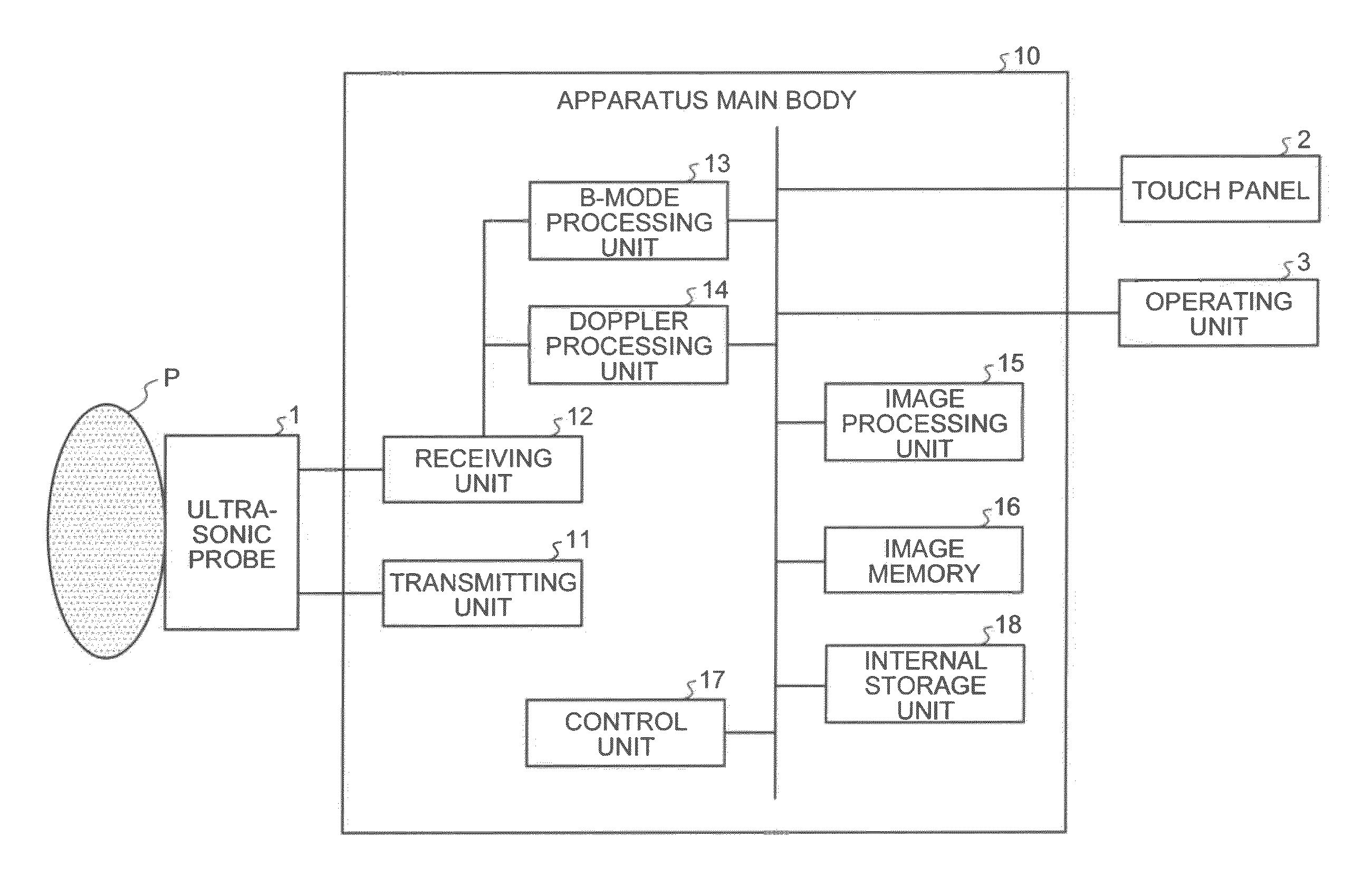
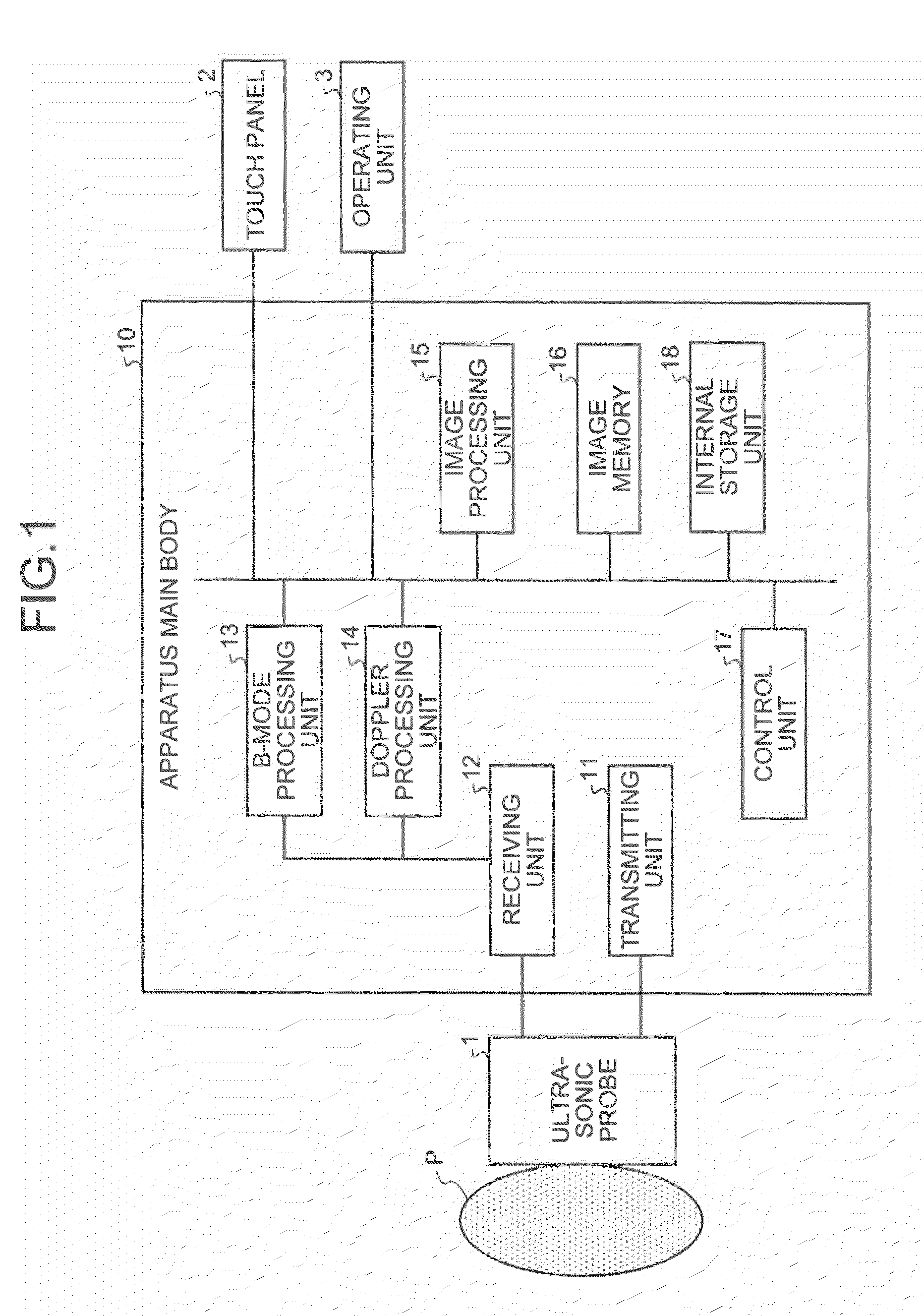
For example, information on the menu for display of moving images and information on the menu for display of still images are provided as the menu information. Here, the menu for moving image display is used when a medical moving image is being displayed. Moreover, the menu for still image display is used when a medical still image is being displayed.

The image storage unit 53b stores therein various medical images obtained from the medical diagnostic imaging apparatus 70 or the medical image storage apparatus 80.

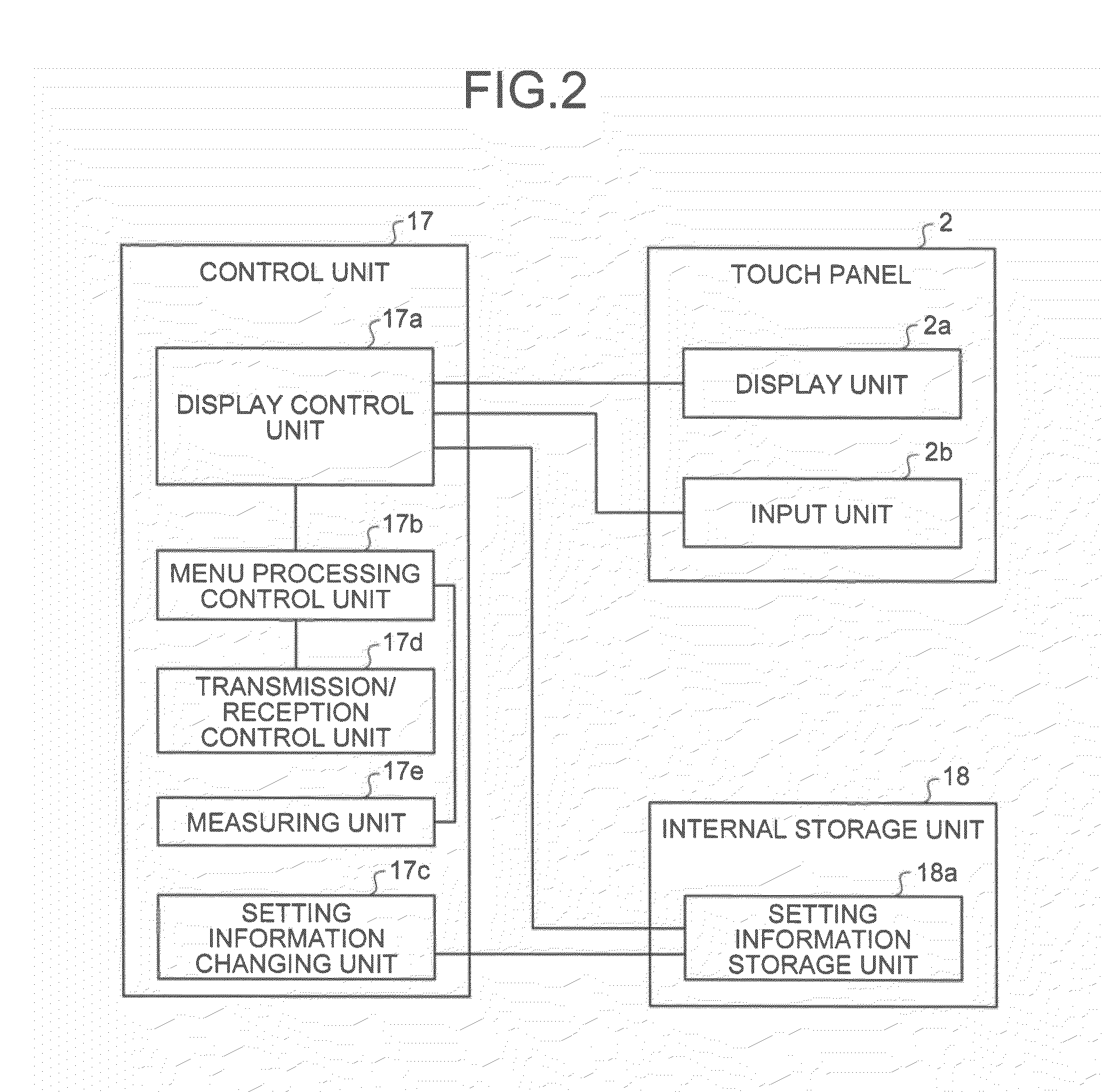
The control unit 54 includes the display control unit 54a, a menu processing control unit 54b, a setting information changing unit 54c, a measuring unit 54e, and an image obtaining unit 54f.

The image obtaining unit 54f obtains a medical image from the medical diagnostic imaging apparatus 70 or the medical image storage apparatus 80 by way of the network 60. For example, the image obtaining unit 54f sends, in response to the operator's request, a medical image obtaining req...


PUM


Abstract

The diagnostic imaging apparatus according to the present embodiments includes a display unit, an input unit, and a display control unit. The display unit includes a display area in which a medical image and a menu in relation to the medical image are displayed. The input unit receives input to the display area. The display control unit displays different menus in the display area in accordance with the input received by the input unit.

Description

CROSS-REFERENCE TO RELATED APPLICATIONSThis application is based upon and claims the benefit of priority from Japanese Patent Application No. 2010-112293, filed on May 14, 2010; and Japanese Patent Application No. 2011-82083, filed on Apr. 1, 2011, the entire contents of all of which are incorporated herein by reference.FIELDEmbodiments described herein relate generally to a diagnostic imaging apparatus, a diagnostic ultrasonic apparatus, and a medical image displaying apparatus.BACKGROUNDA diagnostic ultrasonic apparatus generates an ultrasonic image from a reflection wave signal that is obtained by scanning a subject with an ultrasonic probe. Some of the diagnostic ultrasonic apparatus are provided with a touch panel to display an ultrasonic image and also to receive various operations from an operator. For such diagnostic ultrasonic apparatus incorporating a touch panel, a conventional technology has been known that displays graphics such as switches, icons, and tabs on the monit...

Claims


Application Information

Patent Timeline
IPC IPC(8): A61B8/14
CPCA61B6/463A61B8/465A61B8/463A61B6/465G06F3/0416G06F3/0412G06F3/0484G06F3/04886
Inventor ICHIOKA, KENICHINISHIHARA, KURAMITSUKATAGUCHI, MUNEKI
Owner TOSHIBA MEDICAL SYST CORP
Why Eureka
Try Eureka