Method and apparatus for endoscopic ligament release

a technology of ligament release and endoscopy, applied in the field of endoscopy ligament release surgery, can solve the problems of insanguination of the nerve, numbness, cold feeling, hand and finger pain, etc., and achieve the effect of preventing the re-use of a single-use device and ligaments and other anatomy

Active Publication Date: 2012-01-19
SEG WAY ORTHOPAEDICS +1
View PDF19 Cites 32 Cited by
  • Summary
  • Abstract
  • Description
  • Claims
  • Application Information

AI Technical Summary

Benefits of technology

[0008]The invention pertains to a method, apparatus, and system for cutting anatomic members, such as ligaments, in surgical procedures such as carpal tunnel release, plantar fasciotomy, gastroc release, cubital tunnel release, and tarsal tunnel release. The apparatus includes a retrograde knife and a guide tool for guiding the knife and a scope during surgery. Relevant features include a knife stop for preventing the knife from inadvertently raising out of the k

Problems solved by technology

Carpal tunnel syndrome occurs when the median nerve is squeezed where it passes through the carpal tunnel, thereby

Method used


Image

  • Method and apparatus for endoscopic ligament release
  • Method and apparatus for endoscopic ligament release
  • Method and apparatus for endoscopic ligament release

Examples


first embodiment

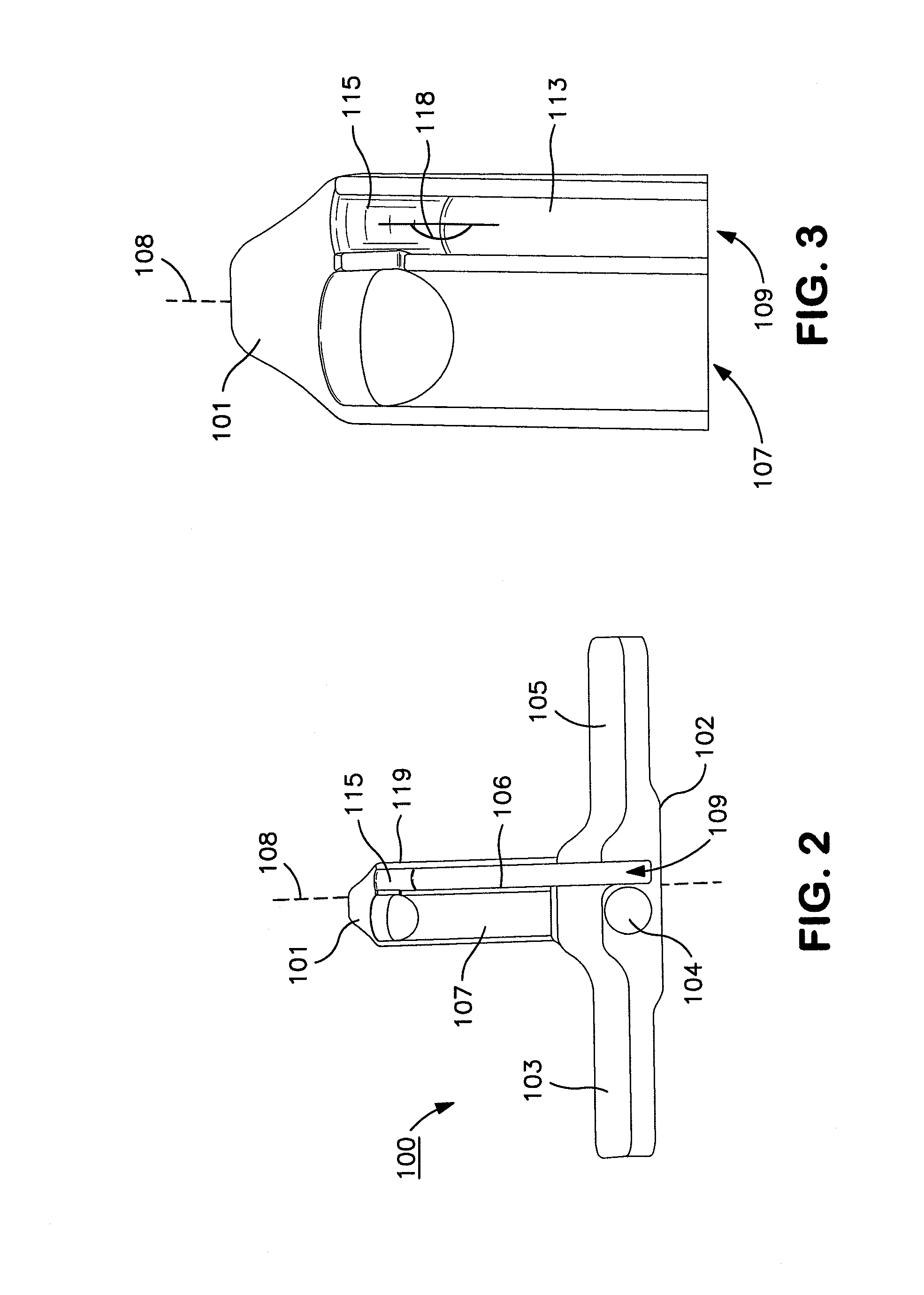
[0025]FIG. 2 is a perspective view looking substantially from the proximal end of a guide member in accordance with the present invention. FIG. 3 is a close up view of the distal end of the guide member of FIG. 2. The guide 100 comprises a longitudinal member 101 and a transverse member 102. The transverse member comprising two wings 103 and 105 extended laterally from the proximal end 104 of the longitudinal member 101. The longitudinal member 101 defines two channels 107 and 109 separated by a partition, such as a ridge 106 in a surface of the longitudinal member for accepting a scope, such as an endoscope or arthroscope, and a knife, respectively.

[0026]One issue of which surgeons must be aware when using carpal tunnel release surgical systems of the type shown in FIGS. 1 and 2 is the fact that the cutting tip of the knife should remain concealed within the knife channel 109 so as not to accidentally engage anatomy when the knife is being inserted distally through the carpal tunne...

second embodiment

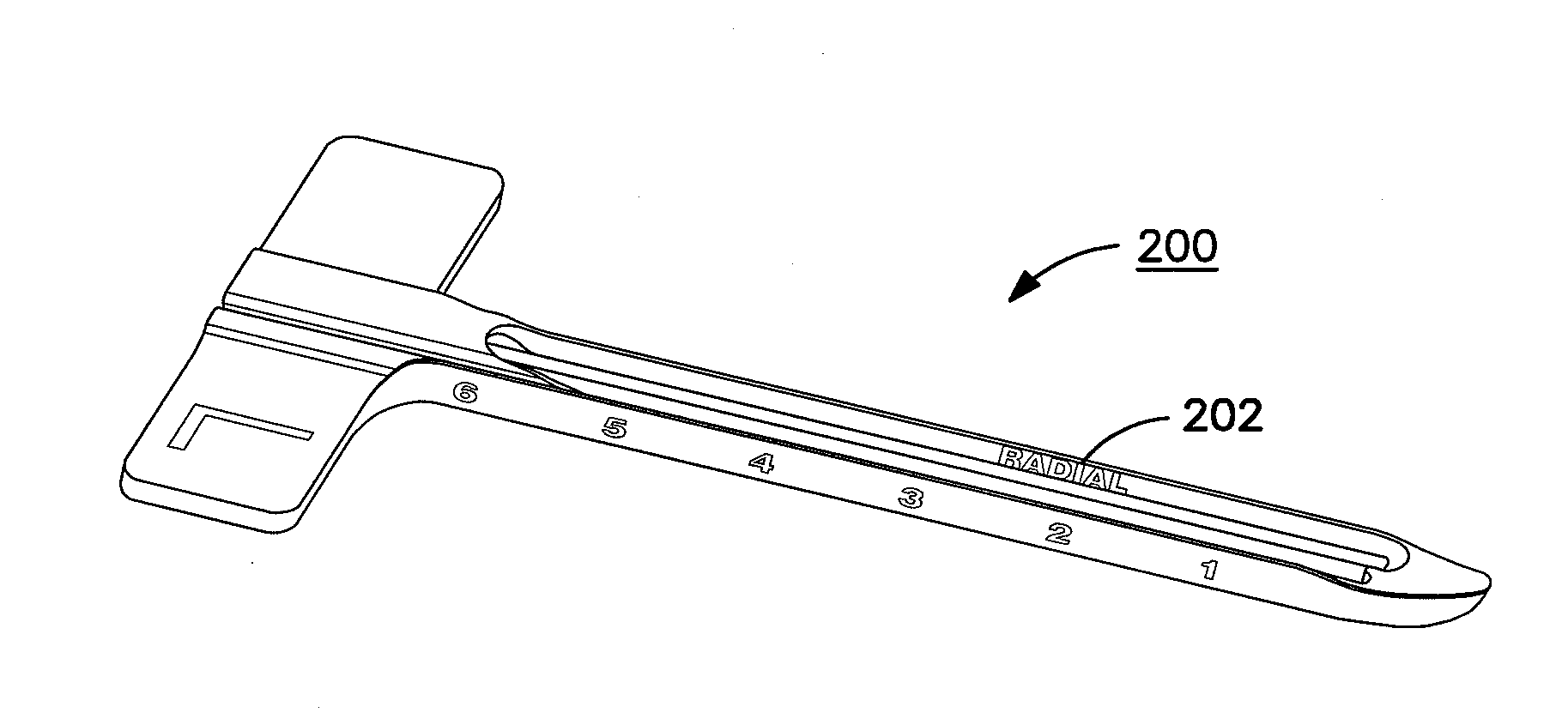
[0030]FIGS. 4A and 4B are right-side and left-side perspective views of the guide 200 illustrating another feature. Particularly, with respect to the human body, medical personnel generally adhere to certain semantic conventions for clarity. For instance, the term “medial” is generally used to describe a direction towards the center of the body, and the term “lateral” is generally used to describe a direction away from the center of the body. However, with respect to the hands, this language is not sufficiently definite because a person can hold one's hand in different orientations, e.g., with the palm facing towards the sky or with the palm facing towards the ground. Accordingly, the terms medial and lateral are indefinite with respect to hands. Hence, with respect to the hand, the terms “ulnar” and “radial” often are used instead. Ulnar refers to the direction from the center of the hand towards the ulnar nerve. The ulnar nerve runs along the length of the hand on the pinky side o...

third embodiment

[0032]FIG. 5 is a perspective view of the distal end of a guide 300 according to a Particularly, as previously described, the distal tip of the guide leads the guide into and through the carpal tunnel. The carpal tunnel is rather tightly packed with flexor tendons and the median nerve. Accordingly, tip 301 is shaped to better assist in dilating the carpal tunnel and spreading the various flexor tendons and radial nerve to create room for the guide to pass through the carpal tunnel.

[0033]More specifically, the distal tip 301 of the guide 300 is prow shaped in order to ease the insertion of the distal tip of the guide into the carpal tunnel and to assist in the dilation of the carpal tunnel and the spreading of the flexor tendons and the medial nerve to allow the longitudinal member 302 of the guide to pass through the carpal tunnel. As can be seen, the distal tip is shaped like the prow of a boat. Particularly, it is pointy without being sharp and it is curved upward slightly.

[0034]...


PUM


Abstract

The invention pertains to a method, apparatus, and system for cutting anatomic members, such as ligaments, in surgical procedures such as carpal tunnel release, plantar fasciotomy, gastroc release, cubital tunnel release, and tarsal tunnel release. The apparatus includes a retrograde knife and a guide tool for guiding the knife and a scope during surgery. Relevant features include a knife stop for preventing the knife from inadvertently raising out of the knife channel, indicators showing the proper orientation for the guide tool, a self dilating tip and channel design on the guide tool, a cover piece and/or pivotable panel system for preventing ligaments and other anatomy from getting caught in the guide tool, a pivot pin and groove system for stabilizing the knife and also assuring that the knife blade is not inadvertently raised out of the channel, and a use indicator for preventing re-use of a single use device.

Description

CROSS REFERENCE TO RELATED APPLICATIONS[0001]This application claims the benefit of U.S. provisional application No. 61 / 364,128 filed on Jul. 14, 2010, which is fully incorporated herein by reference.FIELD OF INVENTION[0002]The invention pertains to methods, apparatus, and systems for performing endoscopic ligament release surgery, such as carpal tunnel release, plantar fasciotomy, gastroc release, cubital tunnel release, and tarsal tunnel release surgery, and similar surgical procedures on anatomic members.BACKGROUND[0003]Carpal tunnel syndrome occurs when the median nerve is squeezed where it passes through the carpal tunnel, thereby causing insanguination of the nerve, which leads to numbness, a cold feeling, and / or pain in the hand and fingers. The carpal tunnel is an anatomic passageway at the base of the wrist through which the median nerve and the flexor tendons for the fingers of the hand pass. It is defined by the carpal bones of the hand and the transverse carpal ligament....

Claims


Application Information

Patent Timeline
IPC IPC(8): A61B17/32
CPCA61B17/320036
Inventor BRIGANTI, RICHARD THOMASMILLER, ALAN B.LEINBERRY, JR., CHARLES F.
Owner SEG WAY ORTHOPAEDICS
Why Eureka
Try Eureka