Flexible neural localization devices and methods

a neural localization and flexible technology, applied in the field of flexible devices, can solve the problems of inability to accurately guide surgical procedures, risk damage to nerve tissue, and system variability, and achieve the effect of reducing the risk of nerve damag

Active Publication Date: 2012-05-17
MIS IP HLDG LLC +1
View PDF6 Cites 46 Cited by
  • Summary
  • Abstract
  • Description
  • Claims
  • Application Information

AI Technical Summary

Benefits of technology

[0053]Any of the device variations described herein may also include electrodes that are concentrically arranged. For example, a ribbon-shaped device may have a top surface with one or more electrode pairs and a bottom surface with one or more electrodes pairs. The electrode pairs may be arranged so that an inner (e.g., −) electrode is surrounded by an outer (+) electrode, or with an inner (+) electrode surrounded by an outer (−) electrode. Concentrically arranged electrodes may provide a limited spread of current compared to bipolar electrode pairs that are not concentric (e.g., arranged adjacently). Surrounding the negative pole with the positive pole of the bipolar pair may therefore help control the current direction.
[0055]Markers may be used to help position the devices appropriately so that the stimulation region may be positioned as desired relative to the target tissue.
[0058]In operation, it may be beneficial to apply force to one or both ends of the device to push the device (and particularly one or more electrodes on the device) “down” (e.g., anteriorly towards the patient's front or ventral side / column). Urging the stimulation region of a ribbon-shaped neural localization device by pushing or pulling the ends may be used as part of any of the methods described herein, but may be particularly helpful when an ambiguous (or no) effect on the nerve is seen when stimulating to help evoke a response. For example, pushing both the distal and proximal end regions of the device when stimulating may help determine if a nerve is between the ribbon device and the target tissue, or if the device is on the opposite side of the ribbon-shaped device from the target tissue.
[0061]In some variations, stimulation from the top and / or bottom of the devices, e.g., by energizing the electrode(s) on the first (top) and second (bottom) surfaces, may not result in a neural response, even when a ramp or range of stimulation intensities are used. To prevent damaging the tissue, the applied stimulation may be kept low (e.g., less than 100 mA, less than 50 mA, less than 30 mA, etc.). In general, it may be desirable to stimulate and confirm that the nerve is on one or the other side of the pathway taken by the device through the tissue by: either pushing or pulling the device from one or both ends (e.g. proximal or distal ends) to urge it towards or away from the target tissue; and / or by changing he polarity of the stimulation; and / or by changing the manner of stimulation. The manner of stimulation may be changed by changing from bipolar to monopolar stimulation. In some variations the manner of stimulation may be changed by changing from simultaneous multipolar (e.g., simultaneous stimulation of multiple electrode connected to a common source, including multiple anodes and multiple cathodes) to sequential multipolar stimulation (e.g., sequentially stimulating each bipolar pair on the same stimulation region). This may allow a greater current density from each bipole pair (or from each monopole, in monopolar configurations), in neural localization devices configured to allow sequential stimulation.

Problems solved by technology

Surgical intervention may require the manipulation of one or more medical devices in close proximity to a nerve or nerves, which may risk damage to the nerve tissue.
Although systems for monitoring neural tissue have been described, these systems are typically imprecise.
Further, many of these systems require large current densities (which may also damage tissue) and may be severely limited in their ability to accurately guide surgical procedures.
Because the conductance of biological tissue may vary between individuals, over time in the same individual, and within different tissue regions of the same individual, it has been particularly difficult to predictably regulate the applied current.
Furthermore, the broadcast fields generated by such systems are typically limited in their ability to spatially resolve nerve location and / or orientation with respect to the medical device.
erves. Although multiple electrodes may be used to stimulate the tissue, the devices, systems and methods described are do not substantially control the broadcast
field. Thus, these systems may be limited by the amount of current applied, and the region over which they can detect
In addition, many surgical manipulations, particularly spinal decompressions, must be performed in difficult to reach regions, and the surgical procedures performed may necessarily need to navigate narrow and tortuous pathways.

Method used


Image

  • Flexible neural localization devices and methods
  • Flexible neural localization devices and methods
  • Flexible neural localization devices and methods

Examples


integrated embodiments

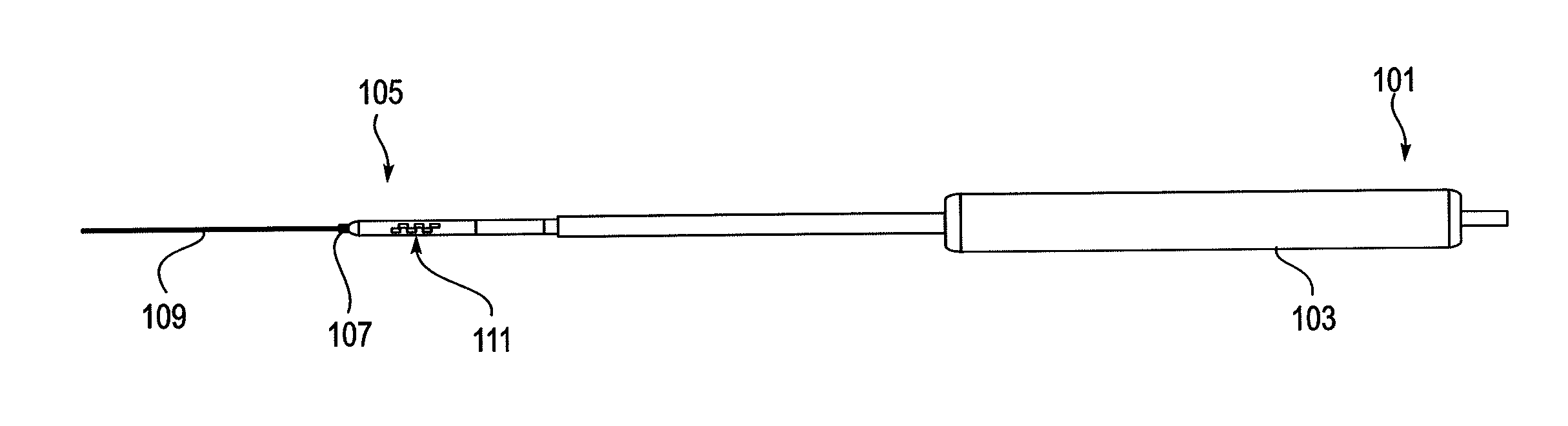
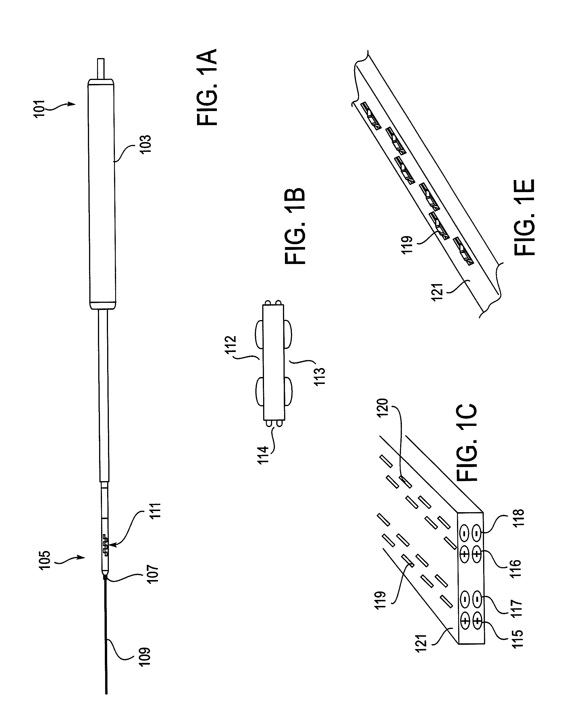
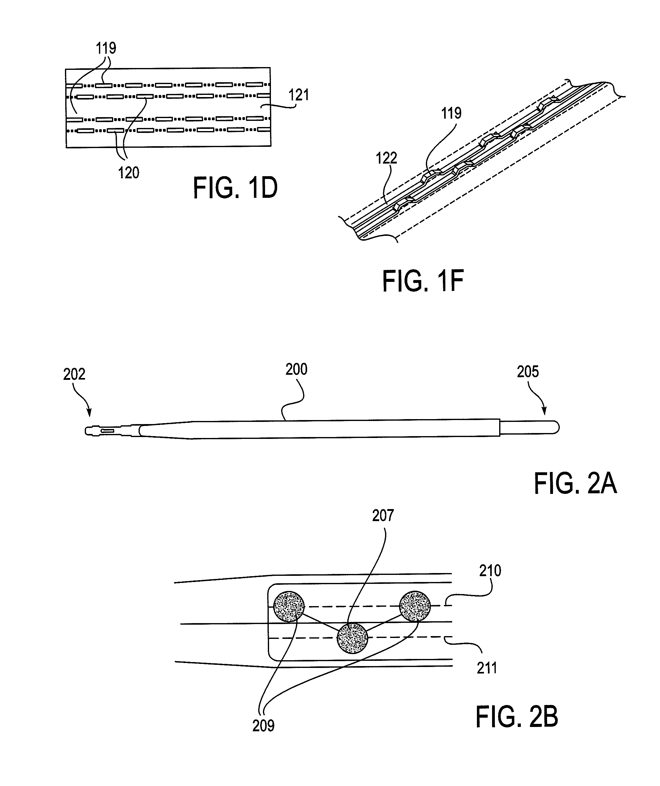
[0226]As mentioned above, the NLR device described herein may couple to one or more tissue modification devices. FIGS. 20A and 20B illustrate two examples of NLR devices 2000 that are configured to couple to tissue modification devices 2001. In FIG. 20A, the tissue modification device 2001 is an elongate flexible tissue removal device that includes tissue modification elements 2003 (e.g., “teeth”) on one side, and has a coupling element at the distal end 2005. The coupling element may be a guidewire coupler, or it may be configured to couple directly to an NLR device 2000. The NLR device 2000 (shown adjacent to the distal end of the tissue modification device in FIG. 20A) includes an opening or cavity into which the tissue modification device may at least partially enter and engage. In other variations, the proximal end of the NLR device is configured to couple to the distal end of the tissue modification device without entering the NLR device.

[0227]The NLR device in this example ma...


PUM


Abstract

Methods for determining if a nerve is nearby a device. The neural stimulation tools described herein are configured to be flexible and low-profile, so that they can be used within body regions that may be tortuous or difficult to reach, such as within a compressed or partially occluded neural foramen. In most cases, these tools described herein are ribbon-shaped and adapted to be manipulated bimanually, applying force to the ends of the devices from separate locations outside of the patient's body. Thus, the distal end region of the device may be configured to couple to the proximal end of a guidewire. One or more surfaces of the devices may include an electrode or multi-polar network of electrodes configured to stimulate only nerves within a predetermined distance of a particular face of the device. Methods of using these devices are described.

Description

CROSS REFERENCE TO RELATED APPLICATIONS[0001]This patent application is a continuation-in-part of U.S. patent application Ser. No. 12 / 724,315, titled “FLEXIBLE NEURAL LOCALIZATION DEVICES AND METHODS”, filed on Mar. 15, 2010, now Publication No. US-2011-0004207-A1; which is a continuation-in-part of U.S. patent application Ser. No. 12 / 504,545, titled “SPINAL ACCESS AND NEURAL LOCALIZATION”, filed on Jul. 16, 2009, now Publication No. US-2010-0010334-A1; which is a divisional of U.S. patent application Ser. No. 11 / 457,416, titled “SPINAL ACCESS AND NEURAL LOCALIZATION”, filed on Jul. 13, 2006, now U.S. Pat. No. 7,578,819; which is a continuation-in-part of U.S. patent application Ser. No. 11 / 251,205, titled “DEVICES AND METHODS FOR TISSUE ACCESS”, filed on Oct. 15, 2005, now U.S. Pat. No. 7,918,849; which claims the benefit of U.S. Provisional Patent Application No. 60 / 681,864, titled “METHODS AND APPARATUS FOR SELECTIVE SURGICAL REMOVAL OF TISSUE”, filed on May 16, 2005. U.S. patent...

Claims


Application Information

Patent Timeline
Patent Type & Authority Applications(United States)
IPC IPC(8): A61B5/05
CPCA61B5/04001A61B2017/143A61B5/1107A61B5/4519A61B5/4893A61B5/6852A61B5/6855A61B17/1671A61B17/1757A61B17/7092A61B17/8897A61B2017/145A61B2562/0209A61B2562/0261A61B2562/043A61B2562/046A61N1/0551A61N1/0553A61B5/0488A61B17/149A61B5/24A61B5/389
Inventor SUN, BENJAMIN KAO-SHINGWALLACE, MICHAEL P.SUMMA, CHRISTOPHER D.
Owner MIS IP HLDG LLC
Why Eureka
Try Eureka