
Producing an image

a technology of producing an image and an organ, which is applied in the field of producing an image of an organ, can solve the problems of difficult success in imaging, poor image quality, and manual operation of known imaging devices, and achieve the effects of improving imaging conditions, easy imaging, and good image quality

Inactive Publication Date: 2012-05-24
OPTOMED OY
View PDF9 Cites 81 Cited by
  • Summary
  • Abstract
  • Description
  • Claims
  • Application Information

AI Technical Summary

Benefits of technology

[0009]The method and system of the invention provide a plurality of advantages. Since the imaging optics can be adapted for each imaging, imaging is easy and image quality good. The camera unit or another part of the system is able to automatically affect image production, whereby the imaging conditions can be improved and / or image processing enhanced for obtaining a successful image.

Problems solved by technology

However, the functions of known imaging devices are manual and intended for one kind of use.
Problems are associated with such solutions.
Although the imaging device is able to produce a fixed image or video image of an organ for the imaging of which the imaging device is not intended, successful imaging is difficult and the quality of the image is not good.

Method used


Image

  • Producing an image
  • Producing an image
  • Producing an image

Examples


Embodiment Construction

[0021]The camera unit of the analytical device may be mainly similar to the solutions disclosed in Finnish published patents FI 107120 and FI 2002 12233, for which reason all properties of a camera unit, known per se, are not described in more detail in the present invention; instead, the focus is on the characteristics of the solution presented that differ from what is known.

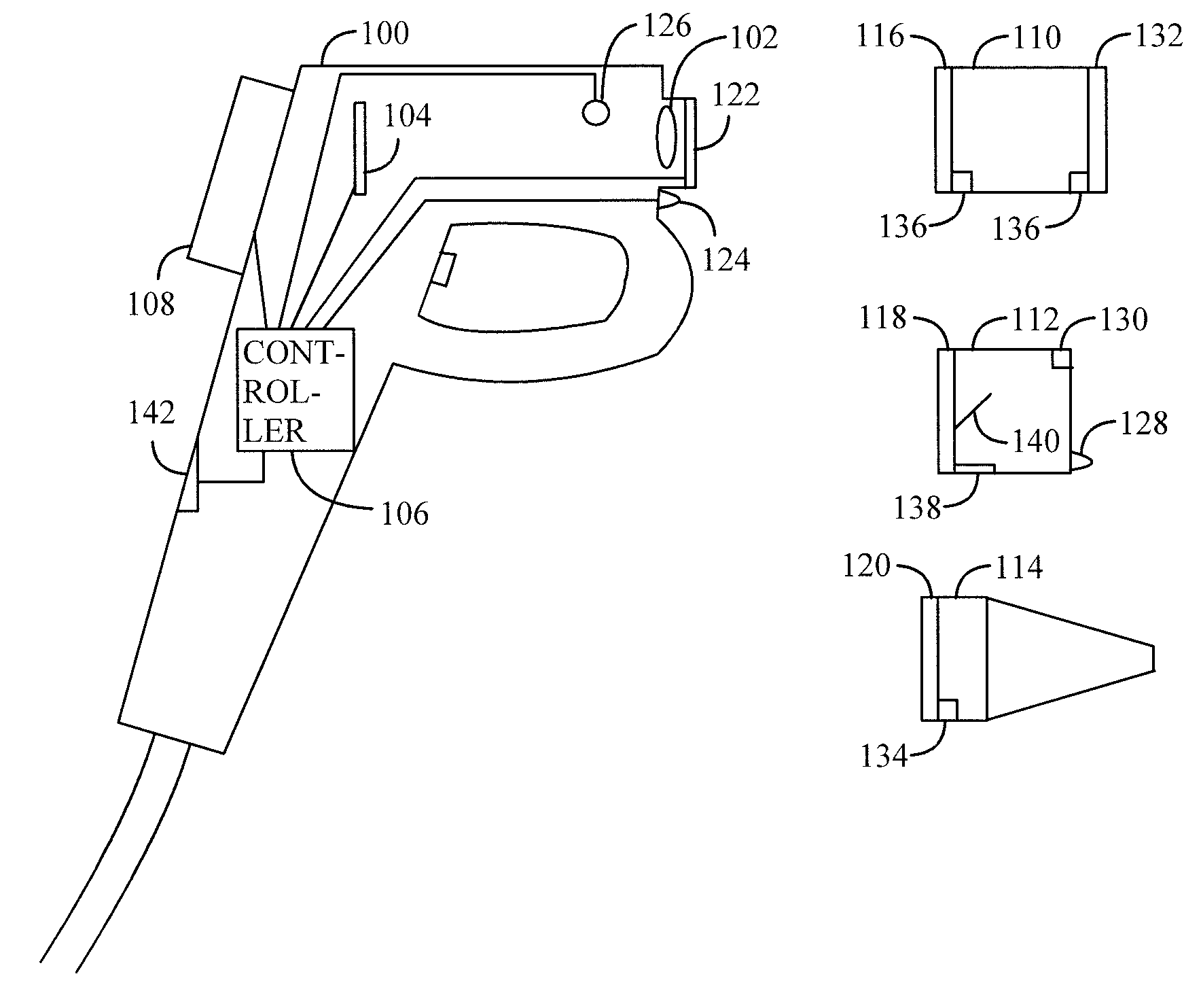
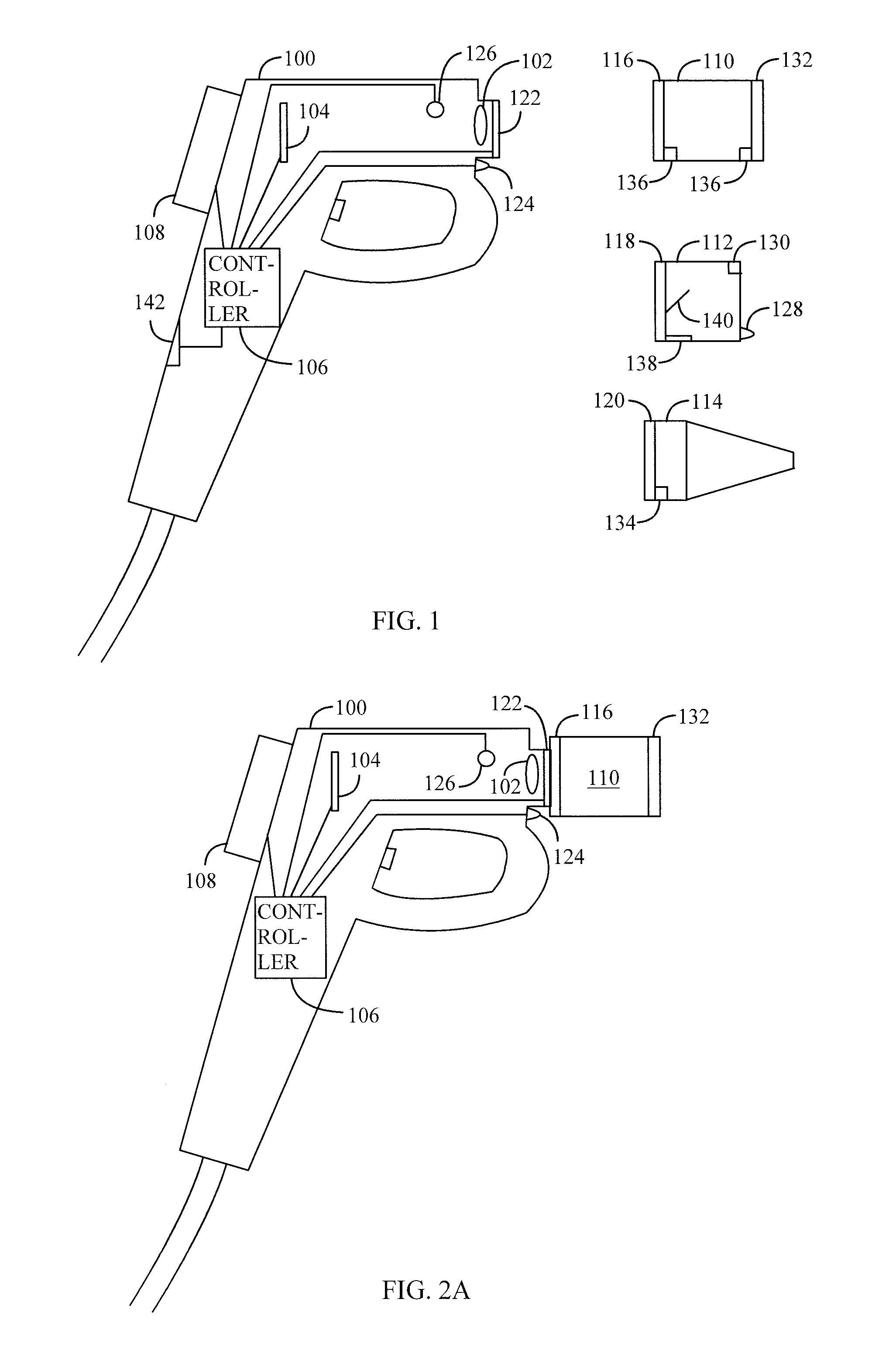
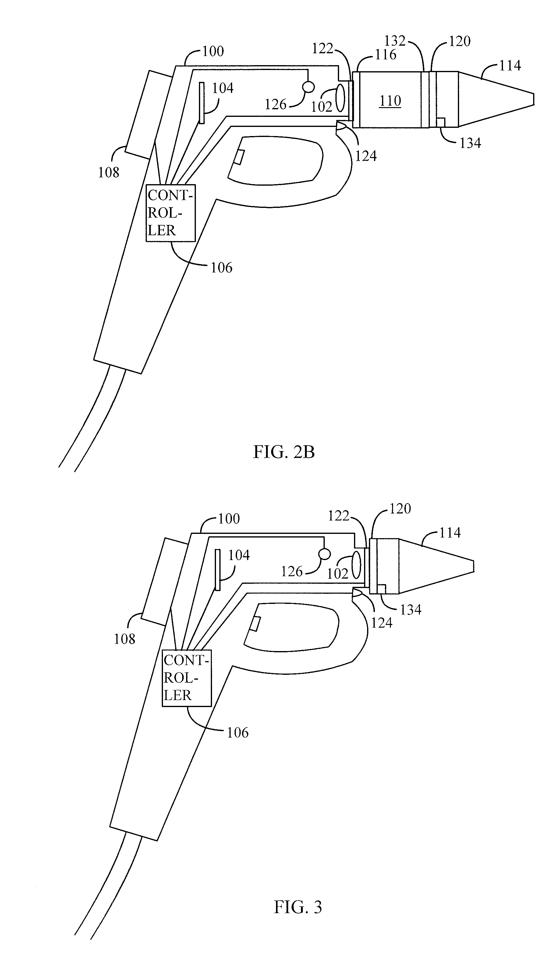
[0022]Let us first study the solution disclosed by means of FIG. 1. In this example, the analytical device is represented by a camera unit 100, which may be a portable camera based on digital technology. The camera unit 100 of the analytical device may comprise an objective 102 for producing an image of an organ to a detector 104 of the camera unit 100. With the analytical device in working order, the detector 104 is able to produce an image in an electronic format of the organ. The image produced by the detector 104 may be input in a controller 106 of the camera unit 100, which controller may comprise a proces...


PUM


Abstract

An optical component is connectable to a camera unit and comprises a data structure including data associated with the optical component. When the optical component is connected to the camera unit, data associated with the optical component is transferred to the camera unit. Image production of an organ by the camera unit is controlled based on the data associated with the optical component.

Description

FIELD[0001]The invention relates to producing an image of an organ by using a camera unit.BACKGROUND[0002]Digital optical instruments may be used as assistance in examining organs, such as the eye, the ear and the skin. For example, a digital image in an electronic format may be produced of an object imaged with a portable camera unit suitable for ophtalmoscopy, otoscopy or dermatoscopy. However, the functions of known imaging devices are manual and intended for one kind of use. This being the case, a device intended for imaging the eye, for example, is not intended for imaging the skin or the ear.[0003]Problems are associated with such solutions. Although the imaging device is able to produce a fixed image or video image of an organ for the imaging of which the imaging device is not intended, successful imaging is difficult and the quality of the image is not good.BRIEF DESCRIPTION[0004]An object of the invention is to provide an improved method and a camera unit for implementing t...

Claims


Application Information

Patent Timeline
Patent Type & Authority Applications(United States)
IPC IPC(8): A61B6/00G03B19/02
CPCA61B1/00009A61B1/00059A61B1/00163A61B2562/08A61B1/227A61B3/14A61B1/04G03B19/02
Inventor POHJANEN, PETRIKOPSALA, SEPPO
Owner OPTOMED OY
Learn More