

Combination therapy with mdm2 and efgr inhibitors
a technology of mdm2 and efgr, which is applied in the field of conjugation therapy with mdm2 and efgr inhibitors, can solve the problem of no approach to overcome such resistan
- Summary
- Abstract
- Description
- Claims
- Application Information
AI Technical Summary
Benefits of technology
Problems solved by technology
Method used
Image

Examples
example 1
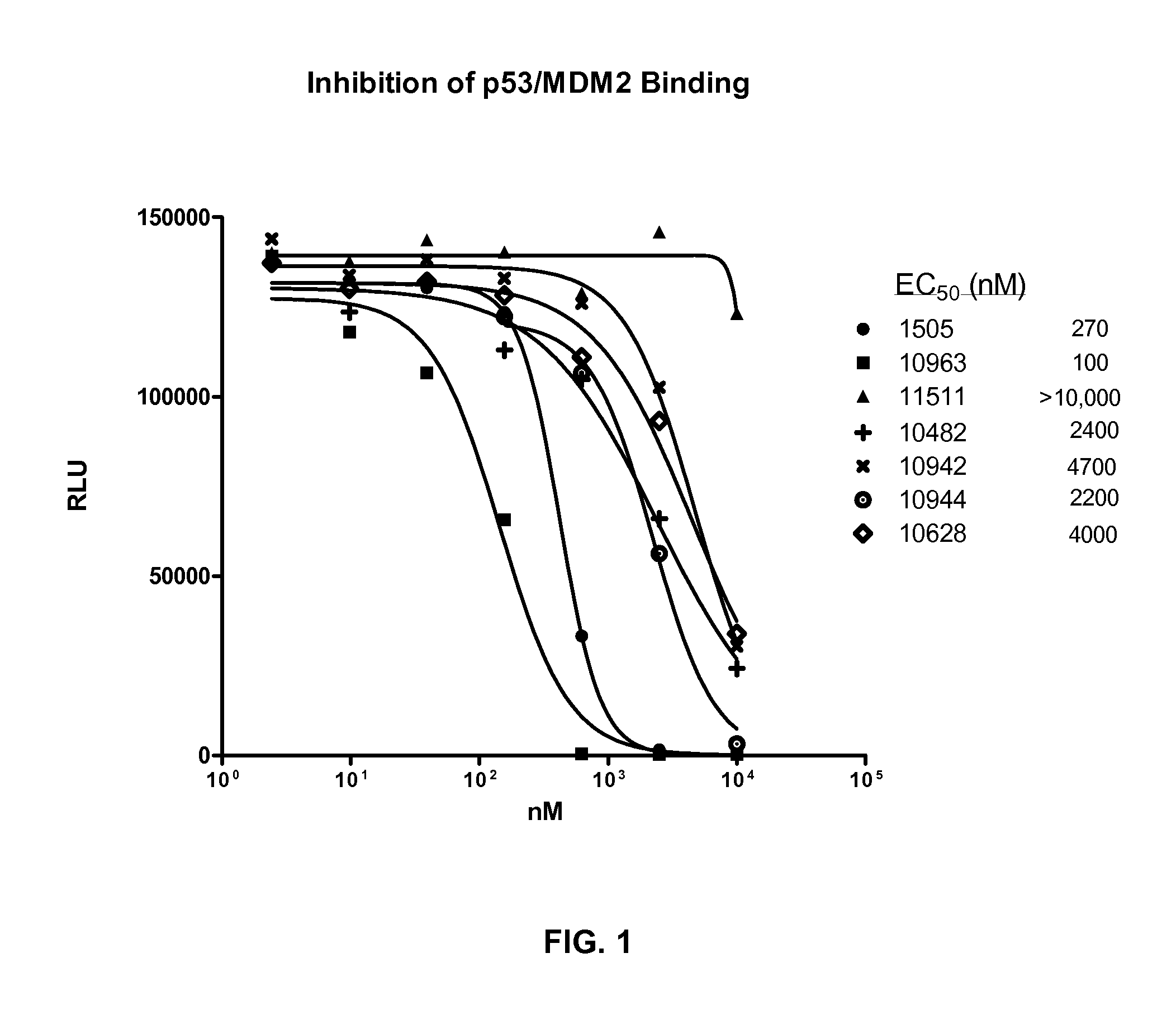
AlphaScreen Measurement of p53 / MDM2 Interaction (Using Truncated MDM2)
[0183]The following example describes an assay that measured the ability of compounds to inhibit the binding of p53 to MDM2 using the AlphaScreen assay technology (PerkinElmer). The following protocol is an adaptation of the method described by H. R. Lawrence et al. (Bioorg. Med. Chem. Lett. 19 (2009) 3756-3759). Recombinant, truncated, human, N-terminal GST-MDM2 (aa 1-150) was obtained from GeneScript. Wild-type, full length human N-terminal 6-his p53 was purchased from SignalChem.
[0184]Resulting in a final reaction volume of 24 μl PBS, 0.1% Tween-20, and 10% glycerol, 30 ng of MDM2 was added, followed by the addition of 1 μl of compound diluted in 100% DMSO that provided a final DMSO concentration of 4%. 30 ng of p53 was then added, mixed, and incubated at room temperature for 1 hour. Glutathione donor beads and Nickel acceptor beads (0.5 μg each; PerkinElmer) were added under subdued lighting conditions to a fi...
example 2
DNA Fragmentation, Measurement of Apoptosis
[0185]The following example describes an assay that measured the ability of compounds to induce DNA fragmentation, an indicator of cell apoptosis, using the Roche Cell Death Detection ELISA kit (Cat #11920 685 001). Methods are according to Example 1 unless otherwise specified.
[0186]Tissue Culture. On day 1, seed A549 cells (10,000 cells / well at 200 μl / well) in tissue culture media (RPMI-1640 with 1% sodium pyruvate, 1% Pen-Strep, 1% L-Glutamine and 10% FBS) were placed in 96-well, tissue culture-treated plates. The plates were allowed to incubate overnight @37° C., 5% CO2. On day 2, the media was removed from the plates and 160 μl media containing 5% FBS was added. 40 μl of media-containing test compound in 100% DMSO (prepared at 5× the dosing concentration) was added to the existing media resulting in a final DMSO concentration of 0.5%. Cells were incubated in the presence of a compound for 24 hrs @37° C., 5% CO2.
[0187]DNA Fragmentation A...
example 3
Proteome Profile, Measurement of Apoptosis
[0189]This example describes an assay that measured the relative expression levels of 35 apoptosis-related proteins in a single sample of whole cell extract. Methods are according to Examples 1-2 unless otherwise specified.
[0190]The protocol and reagents were purchased from R&D systems (Human apoptosis array kit; cat. # ARY009). The kit consisted of an antibody array spotted on nitrocellulose membranes with each specific antibody printed in duplicate. The detection antibodies were biotinylated so they could be used with a streptavidin-HRP conjugate designed for chemiluminescent imaging. The protocol was modified to utilize the LiCor infrared imaging technology by substituting an infrared 680 nm-tagged streptavidin.
[0191]Tissue Culture. A549 NSCLC cells were seeded in 6 well tissue culture plates in RPMI-1640 supplemented with 5% FBS at a density of 1×10⁶ cells / well, 2 ml / well and allowed to incubate overnight at 37° C., 5% CO2, and 85% relat...
PUM
| Property | Measurement | Unit |
|---|---|---|
| Force | aaaaa | aaaaa |
| Volume | aaaaa | aaaaa |
| Volume | aaaaa | aaaaa |
Abstract
Description
Claims
Application Information