Orthodontic Treatment Integrating Optical Scanning and CT Scan Data

a computed tomography and optical scanning technology, applied in tomography, instruments, applications, etc., can solve problems such as gingival recession and mucogingival defects, affecting the health of teeth, and affecting the treatment effect of orthodontic treatment, so as to avoid periodontal defects

Inactive Publication Date: 2012-08-23
GREENBERG SURGICAL TECH
View PDF17 Cites 108 Cited by
  • Summary
  • Abstract
  • Description
  • Claims
  • Application Information

AI Technical Summary

Benefits of technology

[0018]An object of the present invention is...

Problems solved by technology

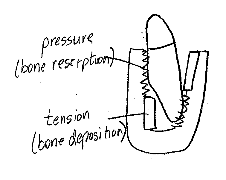
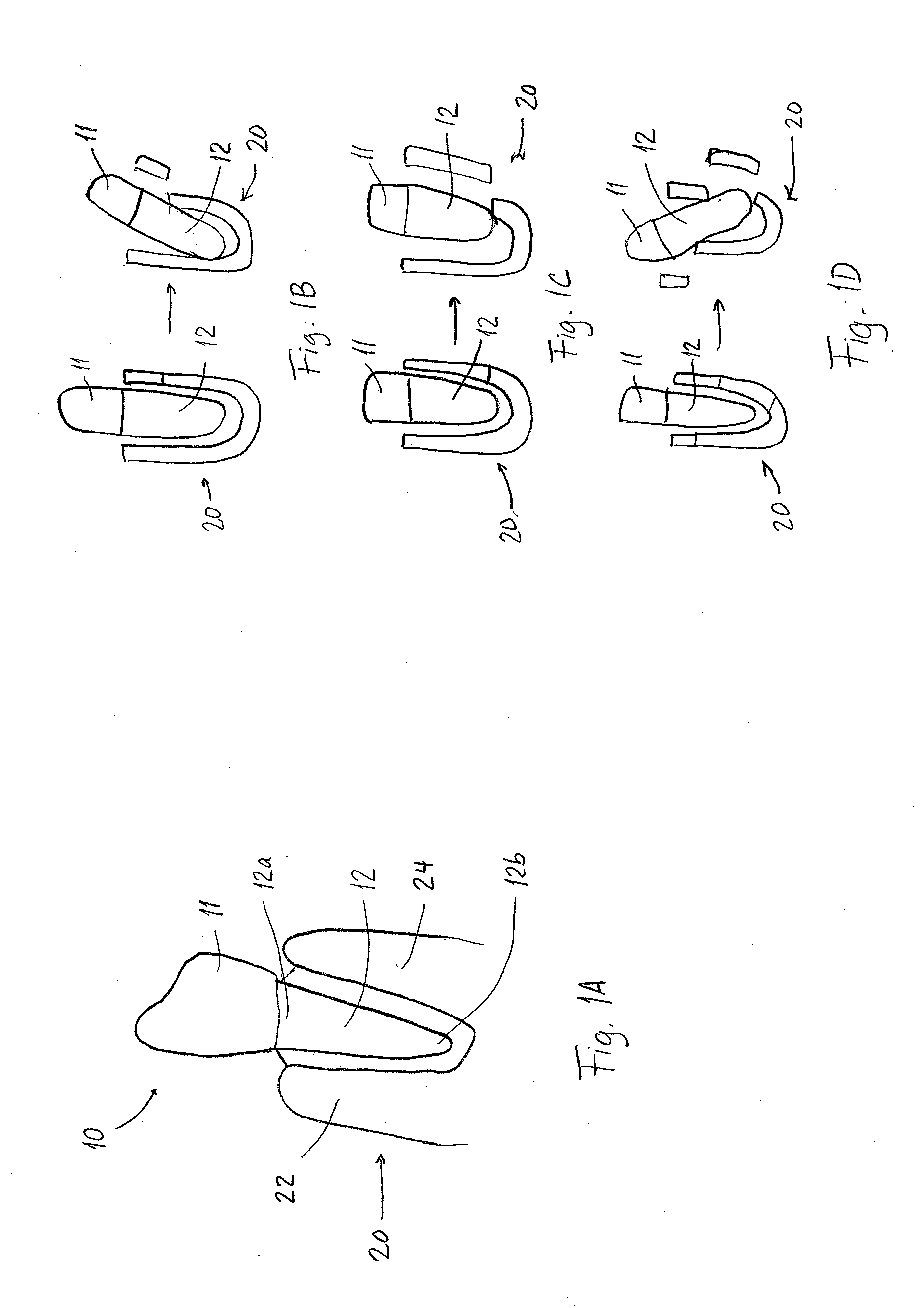
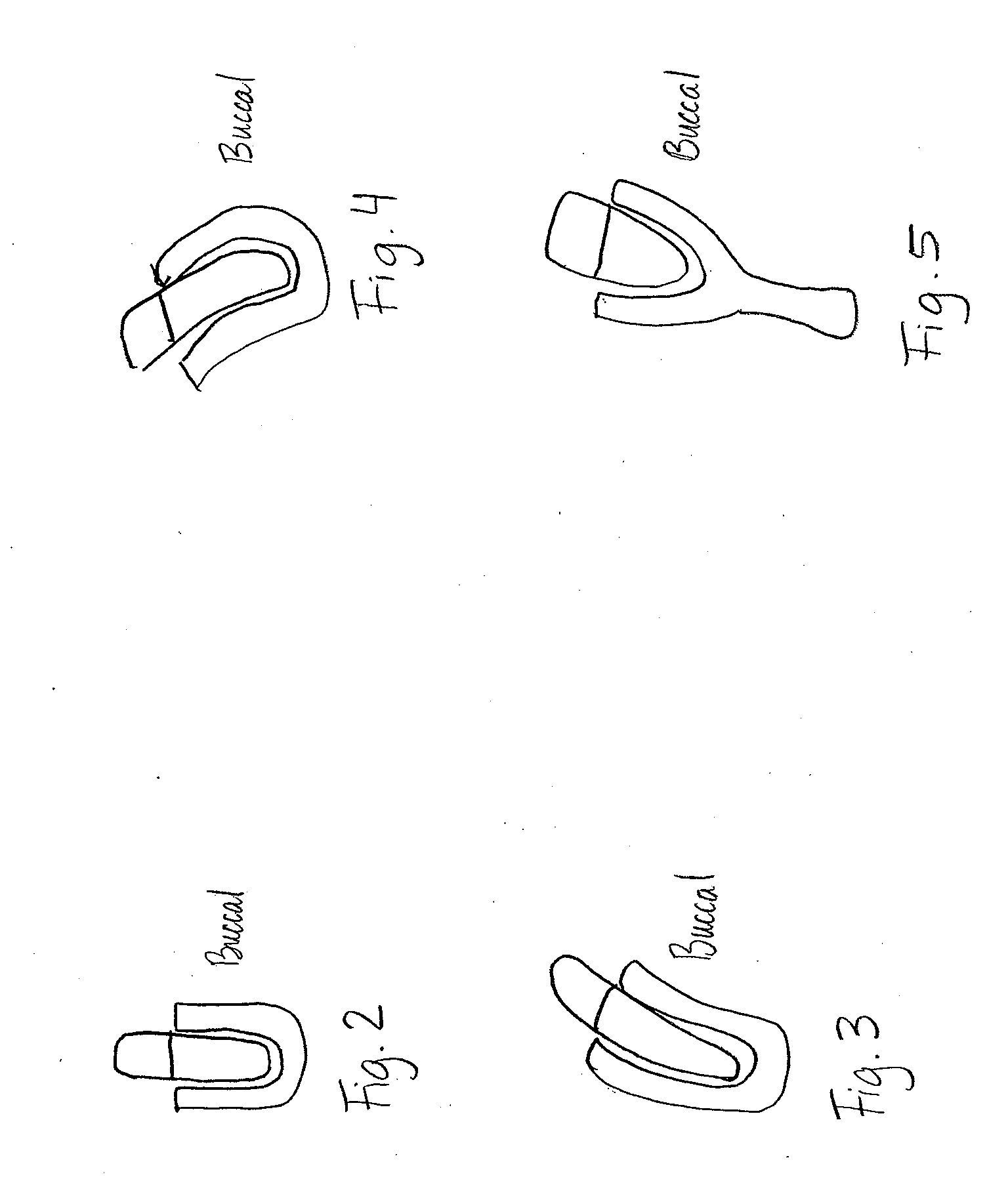
If this movement is excessive or does not properly take into account the alveolar bone morphology, penetration of the bony cortices can occur with resultant gingival recession and mucogingival defects.
A mucogingival defect with its lack of keratinized gingival is less cleansable and less resistant to the effects of bacteria in plaque which causes an inflammatory response that creates a worsening of the periodontal defect and allows the gingival recession and bone dehiscence to deteriorate which jeopardizes the health of the tooth and can cause its premature loss.
This is a greater problem in adult orthodontics than orthodontics in children and adolecesents.
However, if the orthodontic treatment in children and adolescents is not performed correctly, areas of root perforation or penetration with dehiscence can lead to periodontal disease in these patients in their adult years.
FIG. 9 shows that if perforation of the cortical bone occurs on the superior aspect of the root and bone interface, then a root dehiscence is created whereby the loss of periodontal attachment causes gingival recession and if there is complete loss of keratinized gingival which ranges in height from 2-6 or more mm, t...

Method used


Image

  • Orthodontic Treatment Integrating Optical Scanning and CT Scan Data
  • Orthodontic Treatment Integrating Optical Scanning and CT Scan Data
  • Orthodontic Treatment Integrating Optical Scanning and CT Scan Data

Examples


Embodiment Construction

[0048]According to an embodiment of the present invention, a combined scan data showing both a dentition and underlying bone and root structures is created by integrating CT scan data and optical scan data. The integration may be accomplished by one of the following three methods.

[0049]According to a first method of the present invention shown in FIG. 14A, an optical scan of the teeth 1400 and a CT scan of the patient is made 1402 and a superimposition and registration with a 3-dimensional (3D) volume rendering of the dentition based on matching of selected surfaces of the teeth 1404 to produce an artifact corrected image. This is less difficult when there are no dental restorations in the teeth that could create scatter artifact. It may be necessary to use a dental appliance such as an orthodontic bracket as disclosed in U.S. patent application Ser. No. 13 / 299,269, which can be scanned by both CT and optical scan to allow registration of the two datasets, as it contains a radiograp...


PUM


Abstract

A process for creating a dental model to avoid periodontal defects during planned dental work includes obtaining CT scan data and optical scan data of a patient's dentition and integrating the CT scan data and the optical scan data by at least one of surface to surface registration, registration of radiographic markers, and registration of optical markers of known dimensions, to produce a dental model that includes the dentition and underlying bone and root structures. The process then segments anatomic sites of the tooth roots and underlying bone. A plan for the dental work is then generated based on the segmented anatomic sites, whereby the plan avoids periodontal defects based on the knowledge of the anatomic sites of the roots and underlying cortical bones in the dental model.

Description

CROSS-REFERENCE TO RELATED APPLICATIONS[0001]This application claims priority from U.S. Provisional Application Ser. No. 61 / 436,514, filed Jan. 26, 2011, the contents of which are hereby incorporated by reference in its entirety.BACKGROUND OF THE INVENTION[0002]1. Field of the Invention[0003]The present invention relates to the creation of integrated Computed Tomography (CT) and optical scan data for customized orthodontic treatment planning and the production of custom dental models, custom indirect orthodontic brackets, and custom orthodontic aligners that prevent periodontal and mucogingival defects. More specifically, the present invention relates to an integration of a CT scan and an optical scan of dentition derived from an oral cavity or a dental model to prevent the penetration of cortical bone of a jaw in the alveolar region by the tooth root as a consequence of orthodontic movement.[0004]2. Description of the Related Art[0005]In the process of orthodontically applied biome...

Claims


Application Information

Patent Timeline
IPC IPC(8): A61C13/34A61C7/00A61B6/03
CPCA61B5/0088A61B6/032A61B6/14A61C1/084A61B6/5247A61C7/08A61C2201/005A61B6/4417A61C7/002
Inventor GREENBERG, ALEX M.
Owner GREENBERG SURGICAL TECH
Why Eureka
Try Eureka