Medical image processing method, device and system
A medical imaging and processing method technology, applied in the field of three-dimensional imaging, can solve the problems of misoperation of surgery, inaccurate judgment of anatomical parts, etc., and achieve the effect of avoiding inaccurate positioning
- Summary
- Abstract
- Description
- Claims
- Application Information
AI Technical Summary
Problems solved by technology
Method used
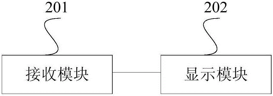
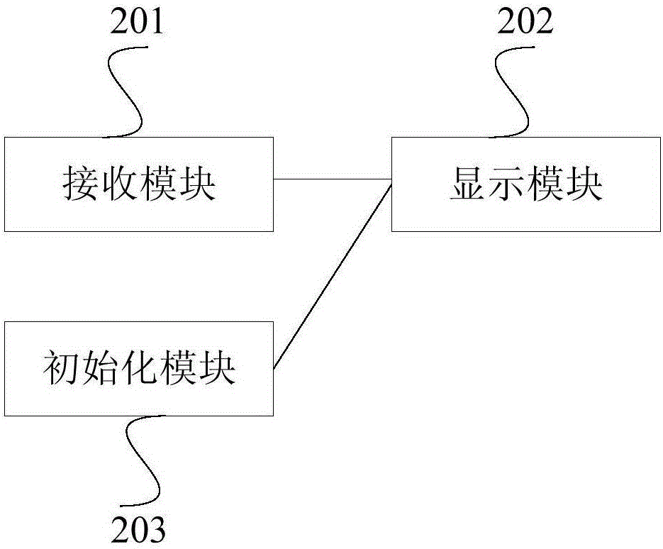
Image

Examples
Embodiment Construction
[0018] The following will clearly and completely describe the technical solutions in the embodiments of the present invention with reference to the accompanying drawings in the embodiments of the present invention. Obviously, the described embodiments are only some, not all, embodiments of the present invention. Based on the embodiments of the present invention, all other embodiments obtained by persons of ordinary skill in the art without making creative efforts belong to the protection scope of the present invention.
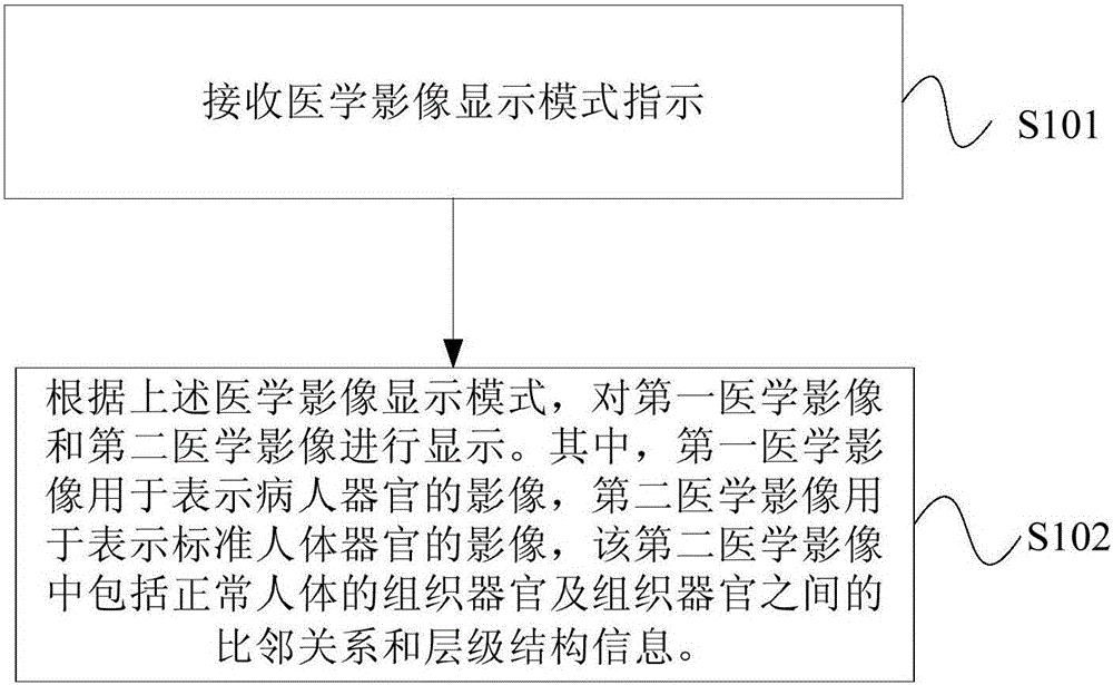
[0019] When doctors use the existing medical image processing system as a reference for preoperative planning and intraoperative navigation, the medical image processing system presents only the patient's medical image information, which is based on CT images, etc. For 3D reconstruction, other information in the original CT image, such as the surface of the human body and the tissue structure from the surface of the human body to tissues and organs, will be rem...
PUM
 Abstract
Description
Claims
Application Information