
Medical device and vascularization method

a medical device and vascularization technology, applied in the field of medical devices and vascularization methods, can solve the problems of not having an established procedure for preventing and curing such ischemic disorders, and achieve the effects of preventing fluid adhesion, facilitating flow, and increasing resistan

Inactive Publication Date: 2012-08-30
TERUMO KK
View PDF4 Cites 25 Cited by
  • Summary
  • Abstract
  • Description
  • Claims
  • Application Information

AI Technical Summary

Benefits of technology

The patent describes a medical device that can be securely fixed to a hole in a bone or other body tissue using a fluid injection method. The device includes a tubular body with a lumen and an expandable body attached to the tubular body. The expandable body expands by the pressure of the fluid injected through the lumen, securing the device to the hole. The device can also include a second tubular body for a second fluid injection. The device can be used to vascularize a hole in a bone by introducing a vascularizing agent and a gel to form a barrier on the inner surface of the hole. The technical effects of this patent include secure fixation of the medical device to the hole, improved fluid injection, and improved vascularization.

Problems solved by technology

This is a serious social problem because aged persons tend to need nursing care once they suffer from femoral fracture.
Unfortunately, there is not any established procedure for preventing and curing such ischemic disorder despite a strong desire.

Method used


Image

  • Medical device and vascularization method
  • Medical device and vascularization method
  • Medical device and vascularization method

Examples


Embodiment Construction

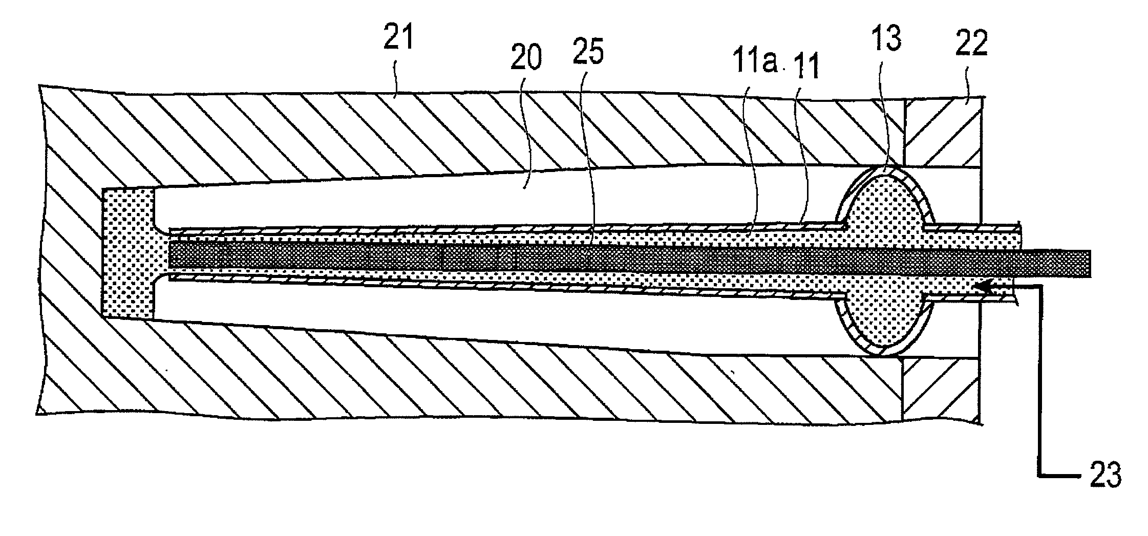
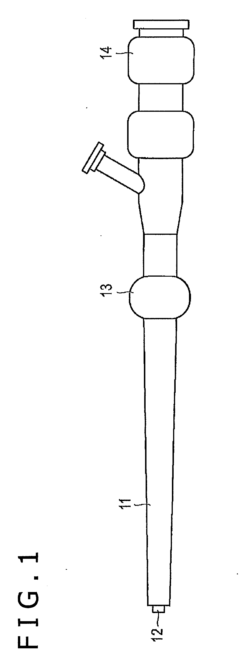
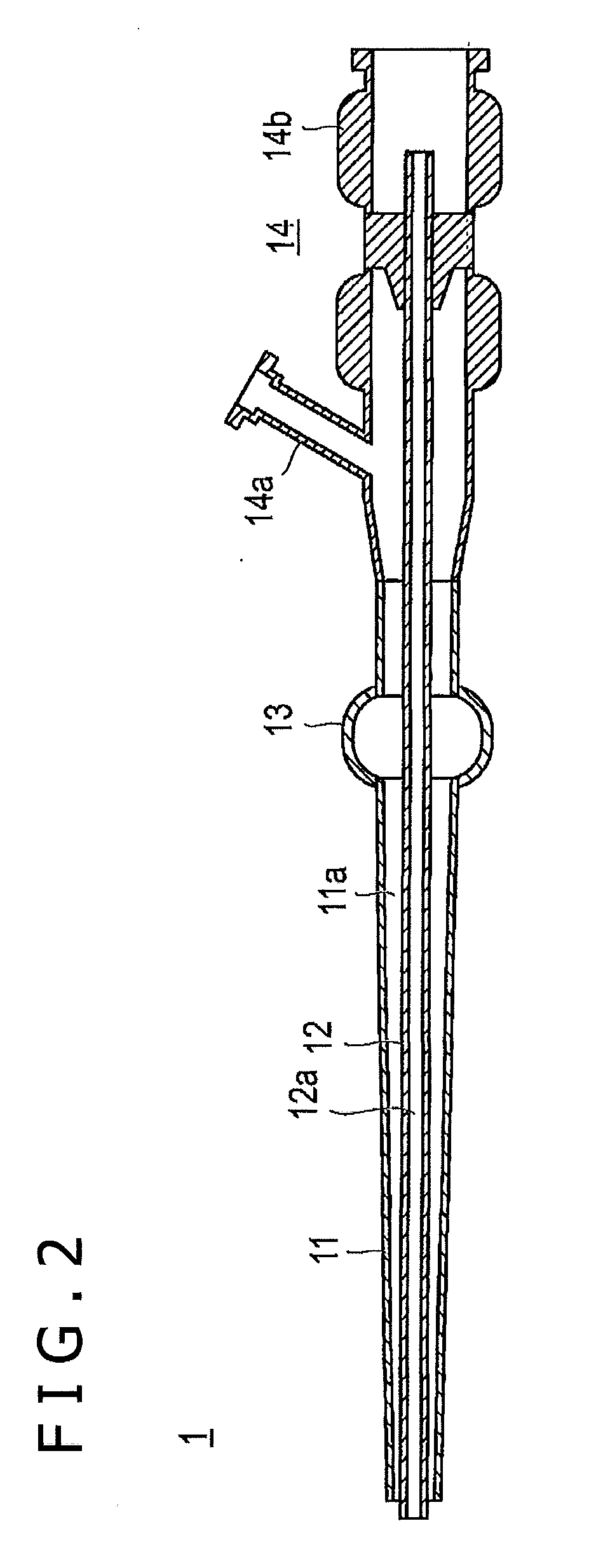
[0038]The following is a description of a first embodiment of the medical device disclosed here. Generally speaking, this first embodiment of the medical device includes a tubular body having a first lumen configured to receive a fluid (gel or liquid) from the opening at one end of the lumen and discharge the fluid from the opening at the other end of the lumen, and an expandable body attached to the tubular body, wherein the first lumen communicates with the space inside the expandable body and the expandable body expands by the internal pressure of the fluid which is injected from the opening at one end and enters the internal space of the expandable body through the first lumen.

[0039]As shown in FIGS. 1 and 2, the medical device 1 according to this first embodiment disclosed by way of example includes a first tubular body 11 having a first lumen 11a, a second tubular body 12 having a second lumen 12a and positioned coaxially in the first lumen 11a, an expandable body 13 arranged ...


PUM


Abstract

A medical device includes a tubular body having a first lumen so designed as to receive a fluid from an opening at one end of the first lumen and discharge the fluid from an opening at the other end of the first lumen, and an expandable body attached to the tubular body. The first lumen communicates with the space inside the expandable body and the expandable body expands by the internal pressure of the fluid which is injected from the opening at one end and enters the space inside the expandable body through the first lumen.

Description

FIELD OF THE INVENTION[0001]The present invention relates to a medical device to inject a fluid into a hole made in the living tissue such as bone and muscle, and a method for vascularization in a hole in a bone.BACKGROUND DISCUSSION[0002]The fracture that occurs most frequently without respect to age is the femoral fracture. Reportedly, 150,000 persons suffer from femoral fracture every year in Japan. This is a serious social problem because aged persons tend to need nursing care once they suffer from femoral fracture.[0003]Known methods for curing femoral fracture include replacement of the bone head with an artificial bone head, bonding the fractured parts with an implant, and conservative treatment. The second method (osteosynthesis) is preferred because it employs the patient's own tissue and is minimally invasive. Osteosynthesis in this case is accomplished by internally fixing the femur.[0004]The implant (such as nail, screw, and pin) used for osteosynthesis of the femur is u...

Claims


Application Information

Patent Timeline
Patent Type & Authority Applications(United States)
IPC IPC(8): A61B17/58
CPCA61B17/3472A61B17/848A61B17/7258A61M25/10A61B2017/561A61B17/8811A61B17/742
Inventor TADA, YUICHINAKAGAWA, YUJIHATA, SUGURUKINOSHITA, YASUSHI
Owner TERUMO KK
Learn More