Renal Cell Carcinoma Biomarkers

a technology of renal cell carcinoma and biomarkers, applied in combinational chemistry, chemical libraries, libraries, etc., can solve the problems of limited treatment options, limited biomarkers, and serious challenges

Inactive Publication Date: 2012-10-04
SIU K W MICHAEL +3
View PDF0 Cites 2 Cited by
  • Summary
  • Abstract
  • Description
  • Claims
  • Application Information

AI Technical Summary

Benefits of technology

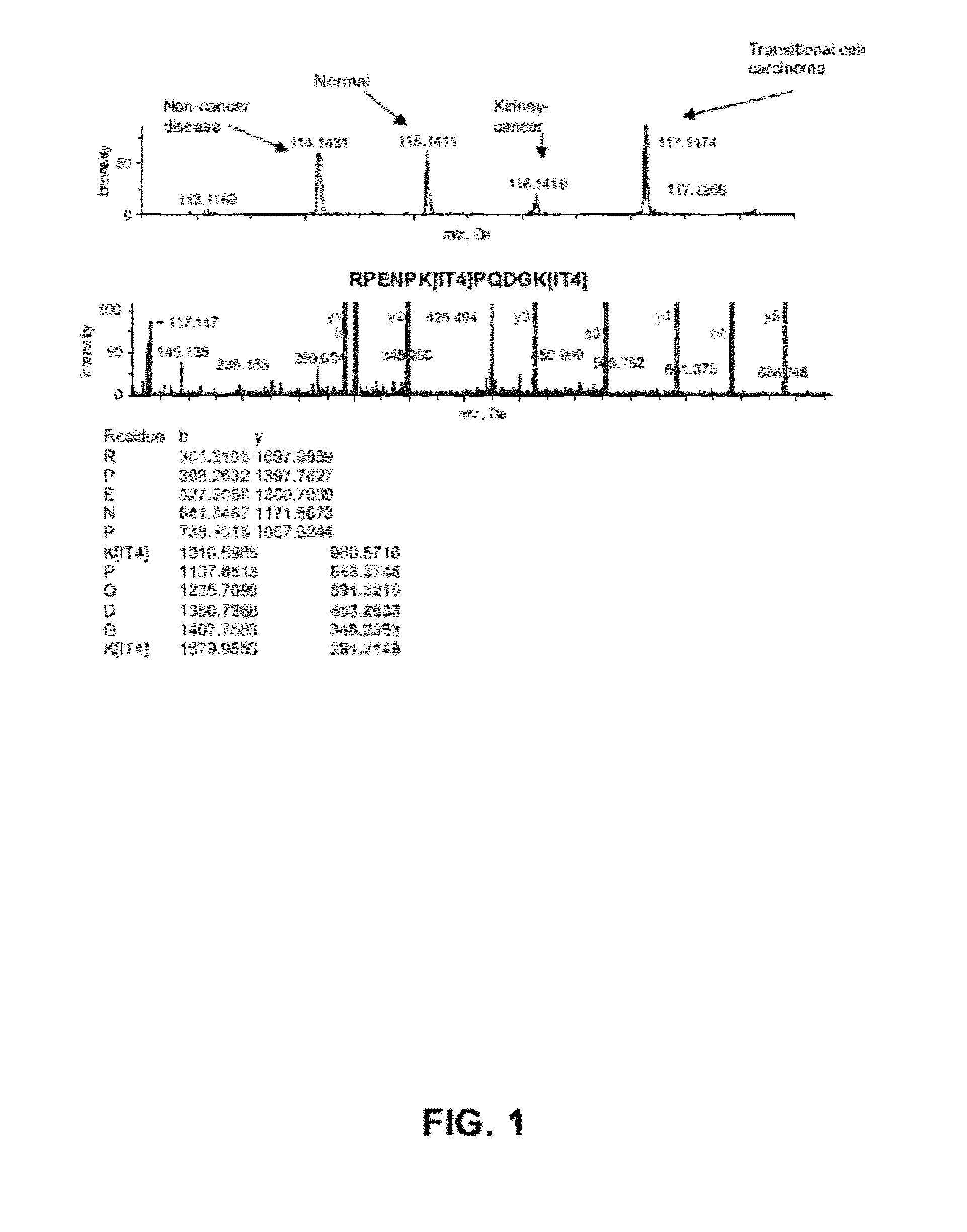
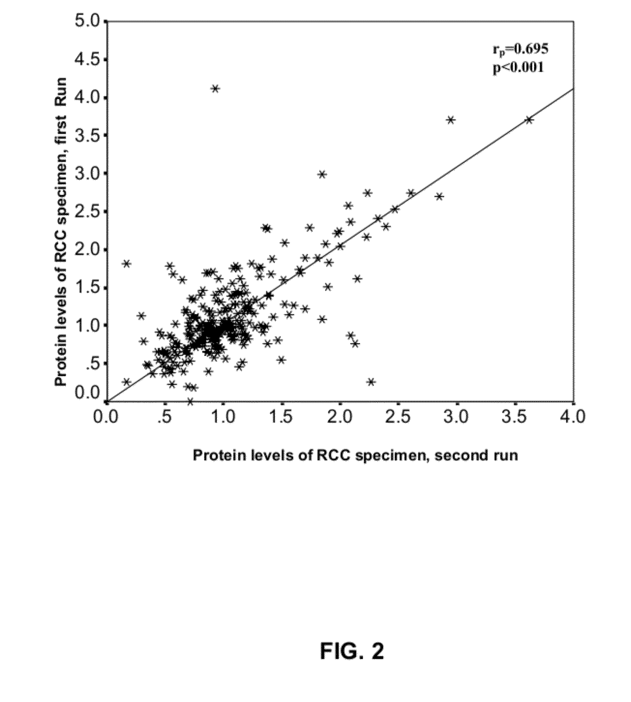
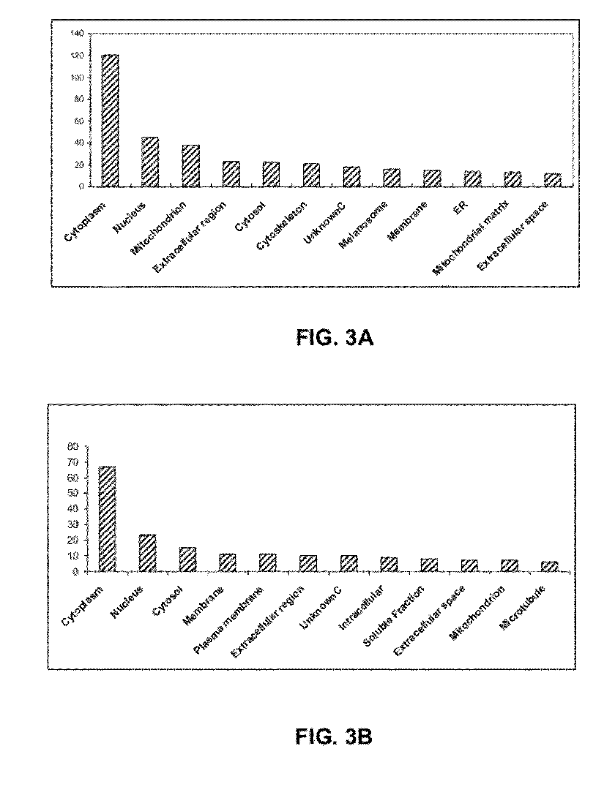
[0010]As described in greater detail herein, the inventors have identified markers associated with renal cell cancer, including renal cell carcinoma. The inventors used multidimensional liquid chromatography-mass spectrometry (LC-MS / MS) for the analysis of biological samples labelled with isobaric mass tags (iTRAQ) to identify proteins that are differentially expressed in renal cell carcinoma (RCC) in relation to adjacent normal counterparts obtained from nephrectomy specimens (control) for cancer biomarker discovery. Two kidney cancer samples were compared against a non-cancerous diseased kidney and normal kidney by online and offline separation. Nine hundred and thirty seven (937) proteins were identified in RCCs, including structural proteins, signalling components, enzymes, receptors, transcription factors and chaperones. Using cutoff values of 1.5 fold for overexpression, and 0.67 fold for underexpression, the inventors were able to identify 168 underexpressed proteins, and 156 proteins that were overexpressed in RCC compared to their normal tissues. These cutoff values were used by the inventors for selecting proteins for further statistical analyses in previous studies (Ralhan et al. 2008) and were found to perform satisfactorily.

Problems solved by technology

The possible existence of non-neoplastic mass lesions can be a serious challenge to the diagnosis of RCC, and there are currently no biomarkers available for the diagnosis of RCC.
Apart from surgery, few treatment options are available because RCC is both chemotherapy- and radiotherapy-resistant.
Heretofore, biomarkers for early detection and follow-up of the disease have not been available, accounting for late diagnosis and subsequent poor prognosis.

Method used


Image

  • Renal Cell Carcinoma Biomarkers
  • Renal Cell Carcinoma Biomarkers
  • Renal Cell Carcinoma Biomarkers

Examples


example 1

Samples and Reagents

[0377]Tumor tissue from patients diagnosed with RCC and their adjacent normal counterparts were obtained from nephrectomy specimens at St. Michael's hospital, after obtaining an informed consent, or from the Ontario Tumor Bank. As RCC is known to arise from the proximal tubules (Pavlovich, Schmidt 2004), the kidney cortex was used as a normal control (Sarto et al., 1997; Shi et al., 2004). The histologic diagnosis for each sample was reconfirmed using microscopic examination of a hematoxylin-and-eosin-stained frozen section of each research tissue block. The tissue from the mirror face of the histologic section was then washed three times in approximately 1 ml of phosphate-buffered saline (PBS) with a cocktail of protease inhibitors, as described previously (1 mM 4-(2-aminoethyl) benzenesulfonyl fluoride, 10 μM leupeptin, 1 μg / ml aprotinin, and 1 μM pepstatin) (1). The washed tissue was then homogenized in 0.5 ml PBS with protease inhibitors, using a handheld hom...

example 2

Strong Cation Exchange (SCX) Separation Conditions

[0379]For the offline 2D LC-MS / MS analysis, each set of labelled samples was first separated by SCX fractionation using an HP1050 high-performance liquid chromatograph (Agilent, Palo Alto, Calif., U.S.) with a 2.1-mm internal diameter (ID)×100-mm length polysulfoethyl A column packed with 5-μm beads with 300 Å pores (The Nest Group, Southborough, Mass.), as previously described (21). A 2.1-mm ID×10-mm length guard column of the same material was fitted immediately upstream of the analytical column. Separation was performed, as previously described (21). Briefly, each pooled sample set was diluted with the loading buffer (15 mM KH2PO4 in 25% acetonitrile, pH 3.0) to a total volume of 2 ml and the pH adjusted to 3.0 with phosphoric acid. Samples were then filtered using a 0.45-μm syringe filter (Millipore, Cambridge, ON, Canada) before loading onto the column. Separation was performed using a linear binary gradient over one hour. Buffe...

example 3

LC-MS / MS Run Conditions

[0381]The inventors used a nanobore LC system from LC Packings (Amsterdam, The Netherlands) consisting of a Famos autosampler and an Ultimate Nano LC system. The LC system was interfaced to an API QSTAR Pulsar-i hybrid quadrupole / time-of-flight (QqTOF) tandem mass spectrometer (Applied Biosystems / MDS Sciex, Foster City, Calif.) equipped with a Protana NanoES ion source (Protana Engineering NS, Odense, Denmark). The spray capillary was a PicoTip SilicaTip emitter with a 10-μm ID tip (New Objective, Woburn, Mass.). The nanobore LC column was 75-μm ID×150-mm length reverse-phase nano capillary column packed in-house with 3 μm C18 beads with 100 Å pores (Kromasil). One μL of sample was injected via the “μL-pick-up” mode. Separation was performed using a binary mobile-phase gradient at a total flow rate of 200 mL / min. For nanospray analysis, the following source conditions were used: a curtain-gas setting of 20 and an ionspray voltage of 1800-3000 V, Q0 declusterin...


PUM

PropertyMeasurementUnit
temperatureaaaaaaaaaa
Kaaaaaaaaaaa

Abstract

The invention provides markers for renal cancer, polynucleotides encoding same, and precursors thereof. The invention also provides methods for detecting, diagnosing, screening for, monitoring, assessing, and treating renal cancers and related disease conditions in a subject. The invention further provides a method of selecting for or assessing efficacy of agents against renal cancer, and a method for assessing renal cancer cell carcinogenic potential of a compound. The invention further provides localization and imaging methods for renal cancers. Also provided are diagnostic compositions and kits for carrying out methods of the invention. In addition, the invention provides therapeutic applications for renal cancers which employ protein renal cancer markers and polynucleotides encoding same, miRNA renal cancer markers and precursors thereof, and binding agents for the markers.

Description

CROSS REFERENCE TO PRIOR APPLICATIONS[0001]This application is a Continuation of PCT Application No. PCT / CA2010 / 000946, filed on Jun. 18, 2010, which claims priority from U.S. Provisional application No. 61 / 213,563, filed Jun. 19, 2009. The disclosures of the aforementioned related applications are incorporated herein by reference in their entirety.FIELD OF THE INVENTION[0002]The invention relates to renal cancer markers, methods for assessing the status of renal cell tissue, and methods for the detection, diagnosis, prediction, and therapy of renal cancer. One aspect of the invention relates to biomarkers of renal cell carcinoma and methods for detection, diagnosis, prediction, and therapy for renal cell carcinoma and related conditions.BACKGROUND OF THE INVENTION[0003]Kidney cancer is a common urologic malignancy that accounts for about 3% of adult malignancies (1a) and causes about 90,000 deaths worldwide annually. Renal cell carcinoma (RCC) is the most common neoplasm in the adu...

Claims


Application Information

Patent Timeline
Patent Type & Authority Applications(United States)
IPC IPC(8): G01N21/64A61K49/00C40B40/06C40B30/04G01N21/76
CPCC12Q1/6886C12Q2600/112C12Q2600/118C12Q2600/136G01N2800/56C12Q2600/178G01N33/57438G01N2500/00G01N2800/52C12Q2600/158A61P35/00
Inventor SIU, K.W. MICHAELDESOUZA, LEROI V.YOUSEF, GEORGE M.ROMASCHIN, ALEXANDER D.
Owner SIU K W MICHAEL
Why Eureka
Try Eureka