
Imaging device, biometric authentication device, electronic equipment
- Summary
- Abstract
- Description
- Claims
- Application Information
AI Technical Summary
Benefits of technology
Problems solved by technology
Method used
Image

Examples
first embodiment
Imaging Device
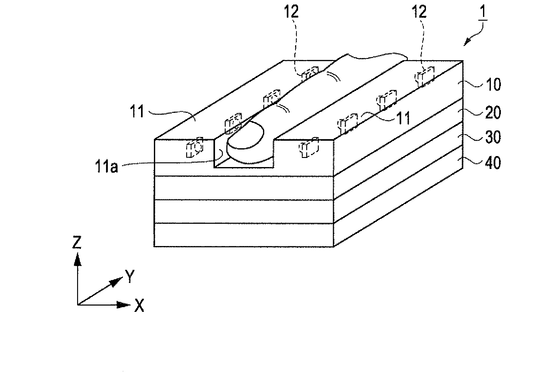
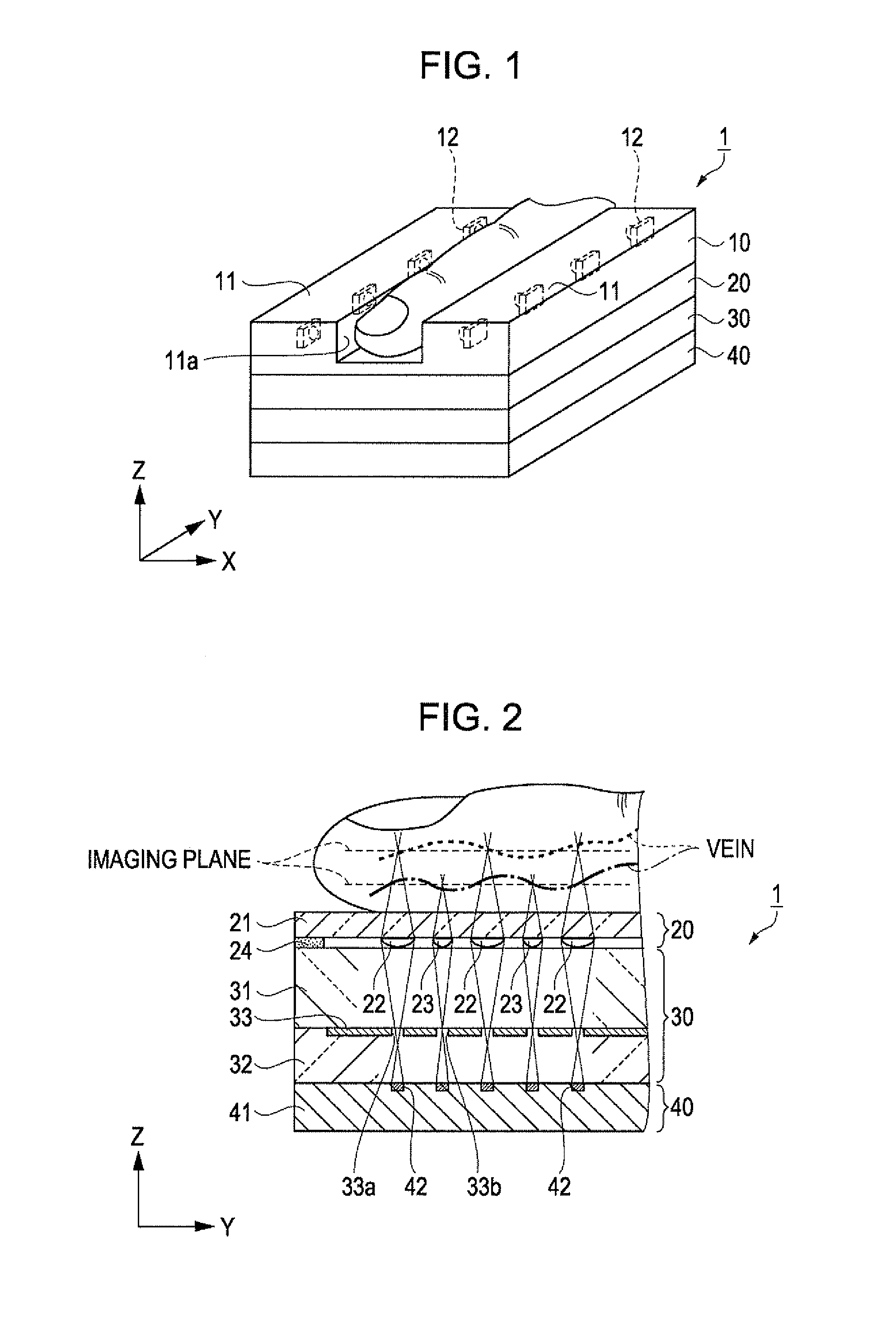
[0036]An imaging device according to a first embodiment is a device that captures an image of finger vein pattern as biological information for identifying a living body. First, the imaging device of this embodiment will be described with reference to FIGS. 1 to 3. FIG. 1 is a schematic perspective view which shows a configuration of an imaging device, and FIGS. 2 and 3 are schematic sectional views which show a configuration of the imaging device. FIG. 2 is a sectional view taken in an extending direction of the vein pattern, and FIG. 3 is a sectional view taken in a direction across (perpendicular to) the extending direction of the vein pattern.
[0037]As shown in FIG. 1, an imaging device 1 according to this embodiment includes, in sequence, a sensor substrate 40 on which a plurality of imaging elements are disposed, a light shielding substrate 30, a lens array 20 on which a plurality of microlenses as a light converging element are disposed so as to converge a light ...
example 1
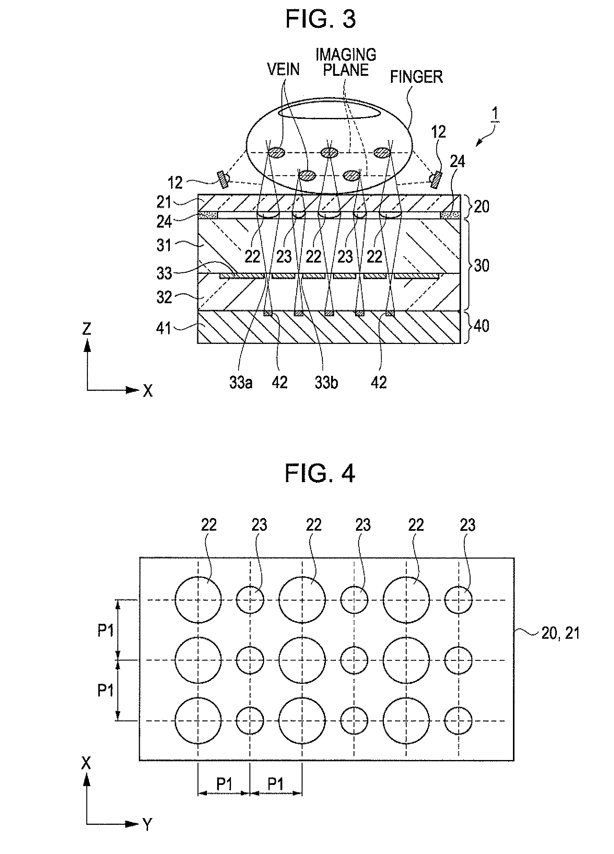
[0048]FIG. 4 is a schematic plan view which shows an arrangement of microlenses of an example 1, and FIGS. 5A to 5D are schematic views which show a process of forming the microlens of the example 1.
[0049]As shown in FIG. 4, the microlenses 22 and 23 having different focal distances are arranged at an equal pitch P1 in the X direction and the Y direction on the transparent substrate body 21 of the lens array 20 of the example 1. The microlenses 23 are positioned between the microlenses 22 in the Y direction, that is, the direction in which the finger extends. With the above arrangement of the microlenses 22 and 23, the vein patterns of the undulated veins running inside the finger as shown in FIG. 2 can be captured in different imaging planes in the Z direction. For example, when the difference between the focal distances of the microlens 22 and the microlens 23 is 100 μm or more, the vein patterns of the undulated veins in different imaging planes can be captured.
[0050]Although var...
example 2
[0061]FIG. 6 is a schematic plan view which shows an arrangement of microlenses of an example 2. The example 2 differs from the example 1 in having a different arrangement of the microlenses. Accordingly, the same configuration is indicated by the same reference numeral and is not described further in detail.
[0062]As shown in FIG. 6, the microlenses 23 having lens diameters smaller than those of the microlenses 22 are arranged at positions between each of the microlenses 22 in the X direction and the Y direction on the transparent substrate body 21 of the lens array 20. In other words, one microlens 23 having a small lens diameter is surrounded by four microlenses 22 each having a large lens diameter. That is, the positioning density of the microlenses 22 and 23 is greater than that of the example 1. As a result, the space on the substrate body 21 is effectively used so that more microlenses 22 and 23 can be positioned. Further, the microlenses 22 are positioned at equal intervals i...
PUM
 Abstract
Description
Claims
Application Information