Rapid screening of monoclonal antibodies
- Summary
- Abstract
- Description
- Claims
- Application Information
AI Technical Summary
Benefits of technology
Problems solved by technology
Method used
Image
Examples
example 1
Binding of Anti-P53 Antibody to Antigen-Coated Microbeads
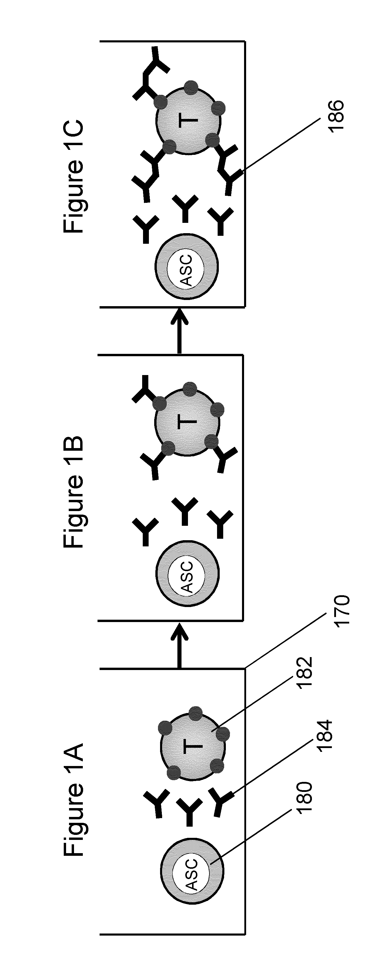
[0124]The purpose of this experiment was to demonstrate a protocol to perform the analysis of antibody secretion by hybridoma cells using polystyrene microbeads as the binding surface. To prove the effectiveness of this method, we suspended p53-coated microbeads with hybridoma cells after having changed the hybridoma supernatant with fresh medium to remove antibodies previously secreted during cell culture. The binding of anti-p53 antibody secreted by hybridoma cells to the microbeads in close proximity was demonstrated.
[0125]Coating of Beads. Polystyrene microbeads having a diameter of 10 μm were suspended in a concentration of 106 beads / mL in borate buffer (0.1M boric acid adjusted to pH 8.5 with 1M NaOH). Microbeads were washed twice in borate buffer and resuspended in 100 μL of borate buffer containing p53 protein in a concentration of 4 μg / mL. The bead suspension was then plated in 4 microwells (25 μL per well) of a 96-we...
example 2
Binding of Antibody Secreted by Murine Hybridoma to Cell Surface Antigen
[0129]The purpose of this experiment was to demonstrate a protocol to perform the analysis of antibody secretion by hybridoma cells and binding of the secreted antibodies to surface antigens on melanoma target cells. To prove the effectiveness of this method, melanoma cells were suspended with hybridoma cells. The hybridoma cells secreted a murine monoclonal antibody capable of reacting with molecules displaying the typical molecular profile of class II MHC antigens. Binding of the secreted antibody to target cells was demonstrated using a hybridoma concentration that is comparable to the equivalent concentration of one cell in a 100 μm microwell in an array of microwells having a 2.25 mm pitch.
[0130]Mouse hybridoma cells were stained with MitoTracker® fluorescent dye (20 nM in PBS for 20 minutes) and resuspended in RPMI+10% FCS. The cell suspension was diluted to 5×10⁴ cells / mL and 50 μL of this suspension were...
example 3
[0132]Activity of NK Cells Against Target 221 Cells
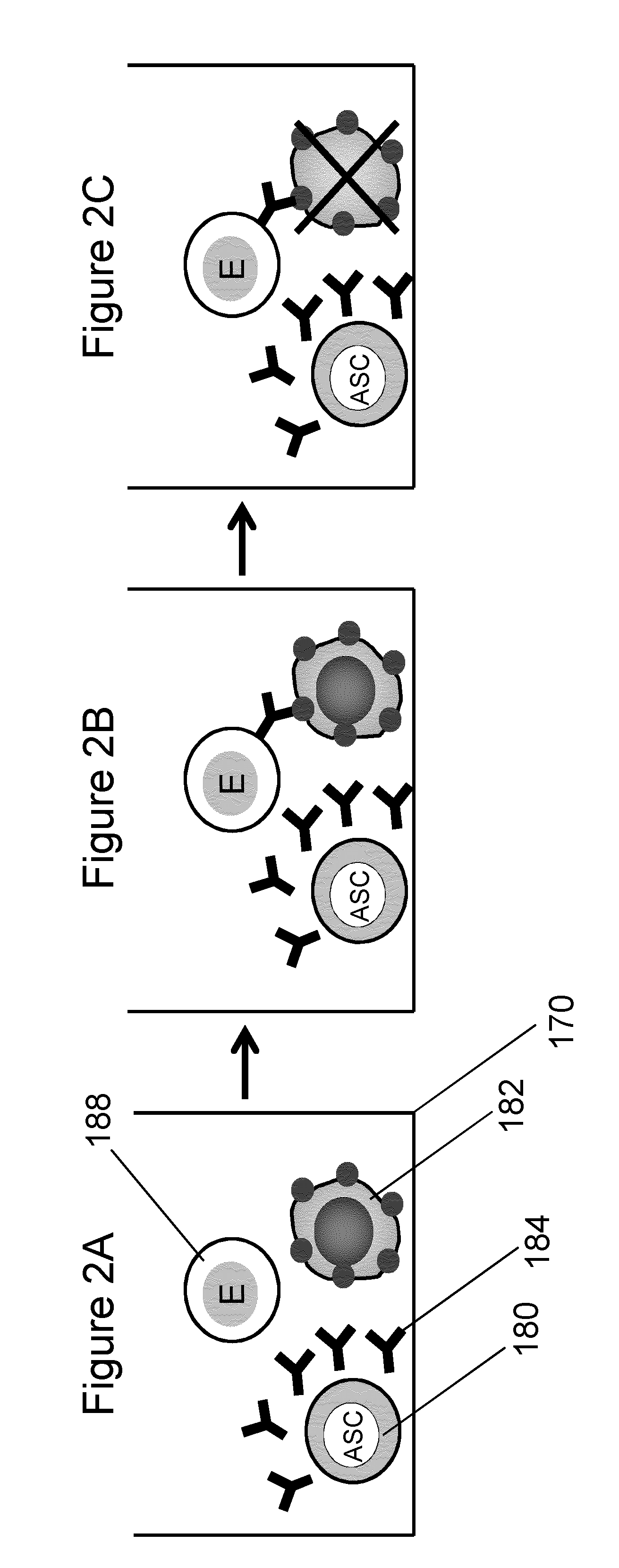
[0133]Analysis of cell-cell interactions at the level of small cell aggregates, sometimes comprising a single target tumor cells was demonstrated in this experiment. The biological system used in this experiment is represented by 221 cells as target cells and by NK and YTS cells working as effector cells. This system has been widely studied with the purpose to understand the immunological mechanisms at the basis of cell-cell interactions and to measure the functional effect of therapeutic monoclonal antibodies in activating the activity of the effector against the target cell. The work of R. Bhat and C. Watzl cited above in this document is an example of the studies conducted on this system.
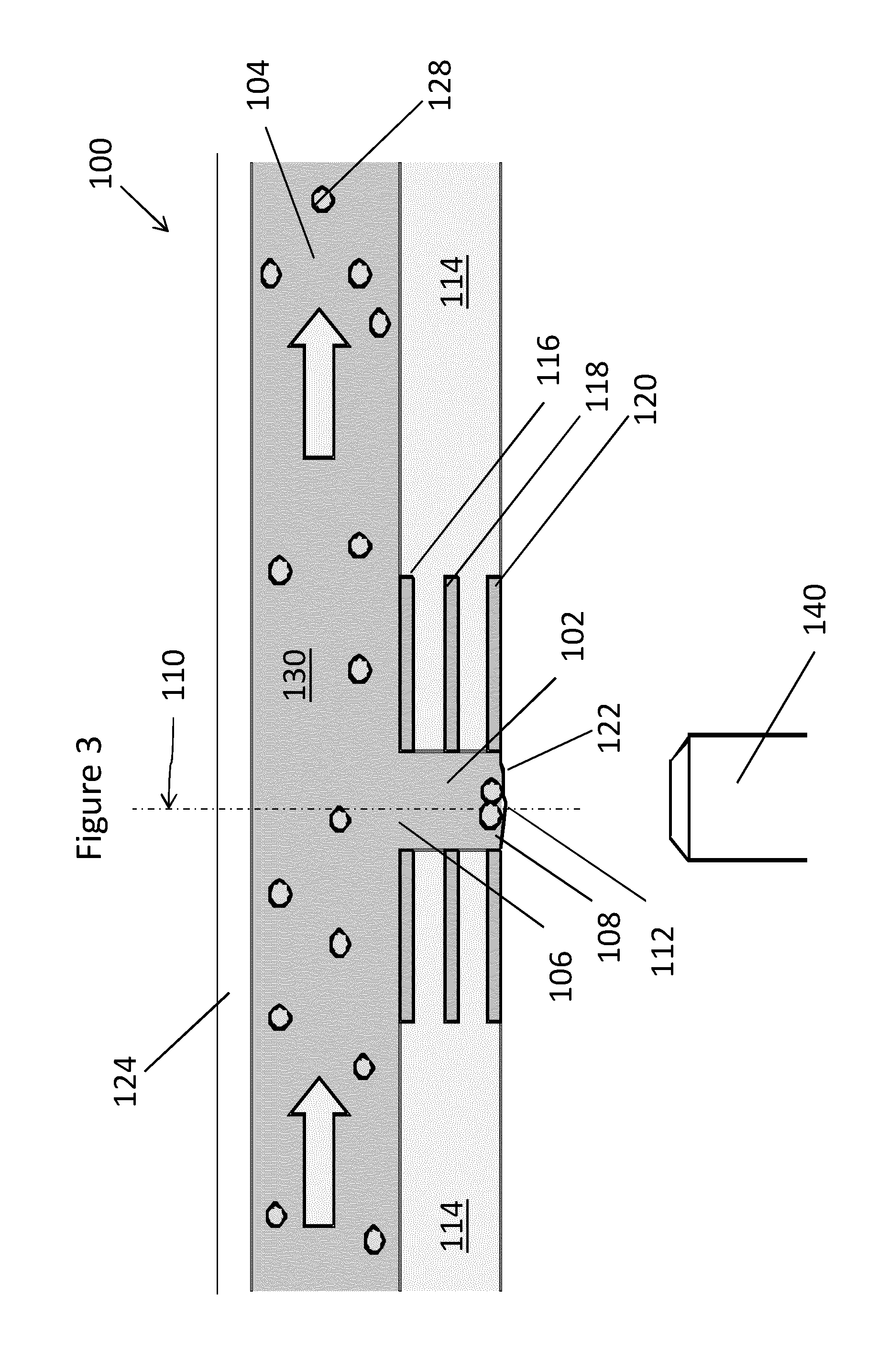
[0134]In the experiment here described the inverted open microwell system was used to isolate the cell aggregates and to monitor the interactions. All cells were cultivated in flasks at 37° C. and 5% CO2 in RPMI medium and for NKL with addition o...
PUM
 Abstract
Description
Claims
Application Information
 - R&D
- Intellectual Property
- Life Sciences
- Materials
- Tech Scout
- Unparalleled Data Quality
- Higher Quality Content
- 60% Fewer Hallucinations
Browse by: Latest US Patents, China's latest patents, Technical Efficacy Thesaurus, Application Domain, Technology Topic, Popular Technical Reports.
© 2025 PatSnap. All rights reserved.Legal|Privacy policy|Modern Slavery Act Transparency Statement|Sitemap|About US| Contact US: help@patsnap.com


