Apparatus and method for breast imaging

a breast and computed tomography technology, applied in the field of medical imaging, can solve the problems of compromising angular range or patient comfort, providing information that is otherwise difficult to ascertain using conventional two-dimensional (2-d) image modalities, and achieving the effect of improving patient comfor

Inactive Publication Date: 2013-10-03
CARESTREAM HEALTH INC
View PDF14 Cites 36 Cited by
  • Summary
  • Abstract
  • Description
  • Claims
  • Application Information

AI Technical Summary

Benefits of technology

[0008]An object of the present invention is to advance the art of 3-D breast imaging and prov

Problems solved by technology

The volume images that can be obtained using tomosynthesis, CBCT, and related techniques have the potential to provide information that can be otherwise difficult to ascertain using conventional two-dimensional (2-D) image modalities.
For breast imaging, the task of acquiring multiple projections from different angles is complicated by the human anatomy itself, which makes it difficult to position the patient in order to obtain the desired field of view for each of the succession of images that are needed.
Proposed solutions for maximizing field of vi

Method used


Image

  • Apparatus and method for breast imaging
  • Apparatus and method for breast imaging
  • Apparatus and method for breast imaging

Examples


Embodiment Construction

[0034]The following is a detailed description of the preferred embodiments of the invention, reference being made to the drawings in which the same reference numerals identify the same elements of structure in each of the several figures.

[0035]Where they are used, the terms “first”, “second”, “third”, and the like, do not necessarily denote any ordinal or priority relation, but are used for more clearly distinguishing one element or time interval from another. For example, there are no fixed “first” or “second” elements in what is described herein; these descriptors are merely used to clearly distinguish one element from another similar element in the context of the present disclosure.

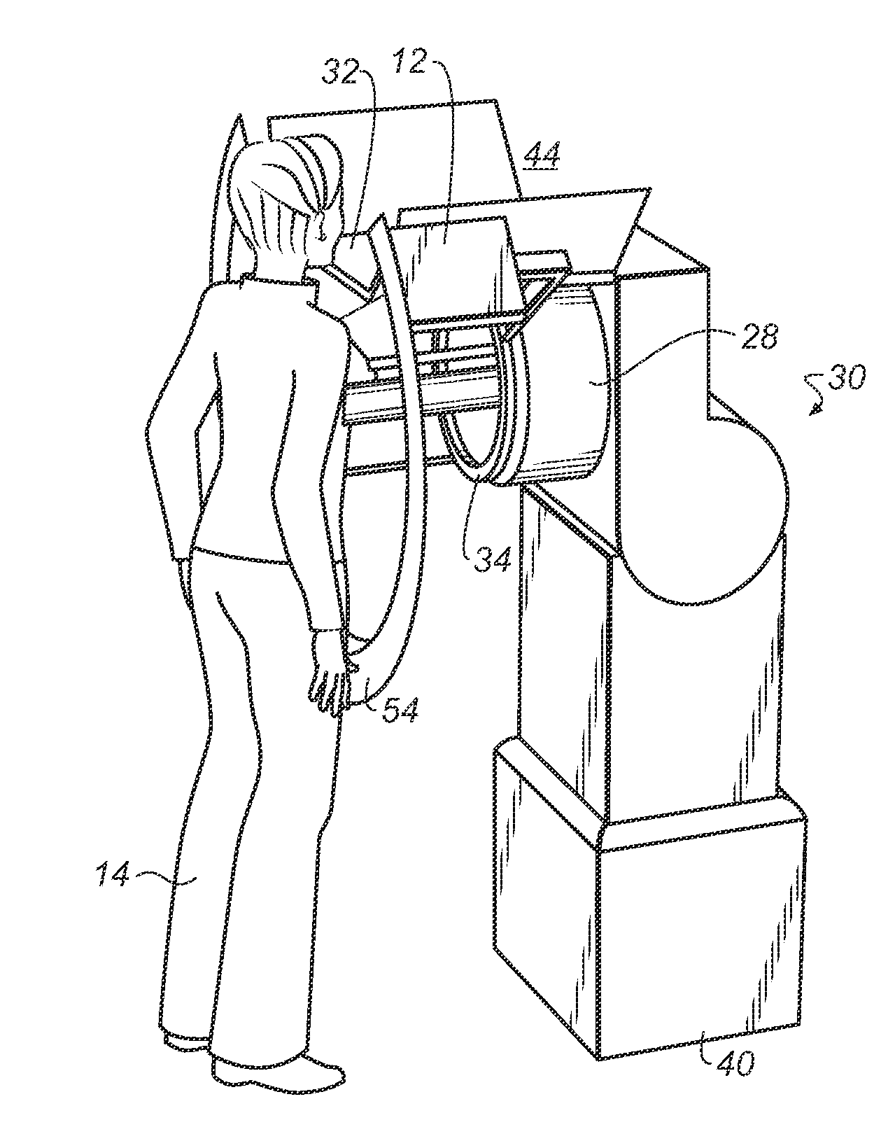
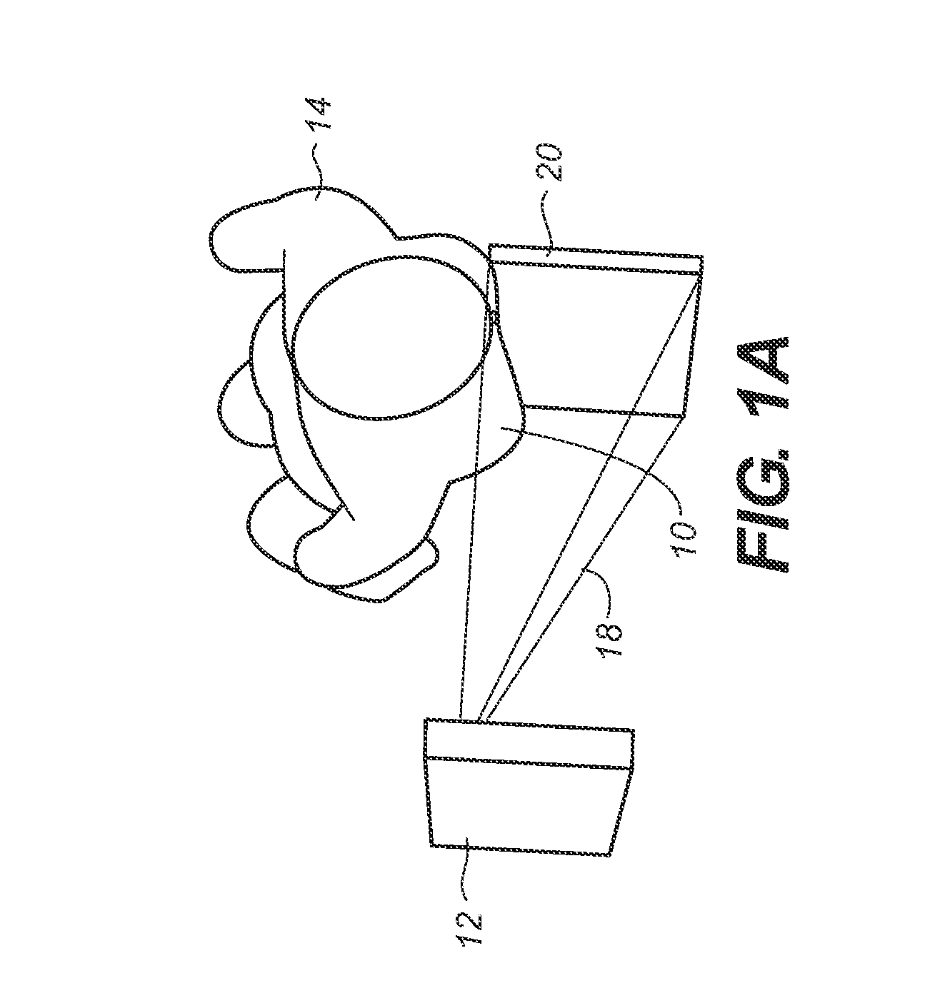
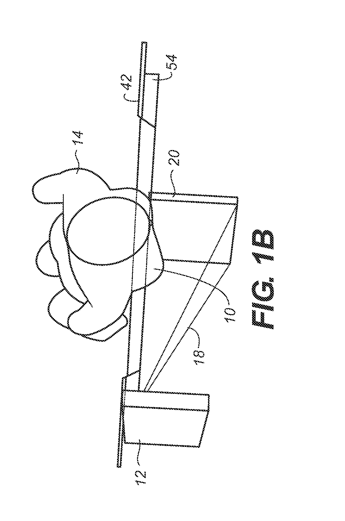
[0036]An issue with existing volume imaging solutions for breast imaging, as noted above, relates to the field of view. Referring to FIG. 1A, there is shown a top view of imaging components for imaging a breast 10 of a patient 14. The imaging system provides a radiation source 12 and a sensor 20, such ...


PUM


Abstract

An apparatus for imaging a breast of a patient has a gantry with a radiation source and a sensor, the source and sensor rotatable in an arcuate orbit about a central axis and within a plane of revolution, wherein the arcuate orbit spans more than 180 degrees and less than 360 degrees, and wherein the gantry has a gantry cover that is disposed to be in contact with at least the chest wall of the patient. The gantry cover has a central opening about the central axis for insertion of the breast that is to be imaged and a peripheral cutout portion that defines the end-points of the arcuate orbit and that provides a space for positioning a portion of the patient's anatomy.

Description

FIELD OF THE INVENTION[0001]The invention relates generally to the field of medical imaging and more particularly to apparatus and methods for cone beam computed tomography of the breast.BACKGROUND OF THE INVENTION[0002]Tomographic volume imaging techniques provide enhanced information over conventional two-dimensional mammography and can help to provide data that lead to early diagnosis and treatment of breast cancer. Cone beam computed tomography (CBCT), for example, is acknowledged to have particular advantages for providing volume images that can be suitable for 3-D mammography. The volume images that can be obtained using tomosynthesis, CBCT, and related techniques have the potential to provide information that can be otherwise difficult to ascertain using conventional two-dimensional (2-D) image modalities.[0003]Volume imaging of a subject requires coordinated movement of the radiation source and sensor, with one or both moving over at least a partial revolution about the subj...

Claims


Application Information

Patent Timeline
IPC IPC(8): A61B6/04
CPCA61B6/025A61B6/502A61B6/0414
Inventor PACKARD, NATHAN J.RICHARD, SAMUELYANG, DONG
Owner CARESTREAM HEALTH INC
Why Eureka
Try Eureka