
Devices and Methods for Using Controlled Bubble Cloud Cavitation in Fractionating Urinary Stones

a technology of controlled bubble cloud cavitation and urinary stones, which is applied in the field of ultrasonic treatment of urinary stones, can solve the problems of reducing efficiency, affecting the treatment effect, so as to suppress cavitation and suppress cavitation

Inactive Publication Date: 2013-11-14
RGT UNIV OF MICHIGAN +1
View PDF3 Cites 27 Cited by
  • Summary
  • Abstract
  • Description
  • Claims
  • Application Information

AI Technical Summary

Benefits of technology

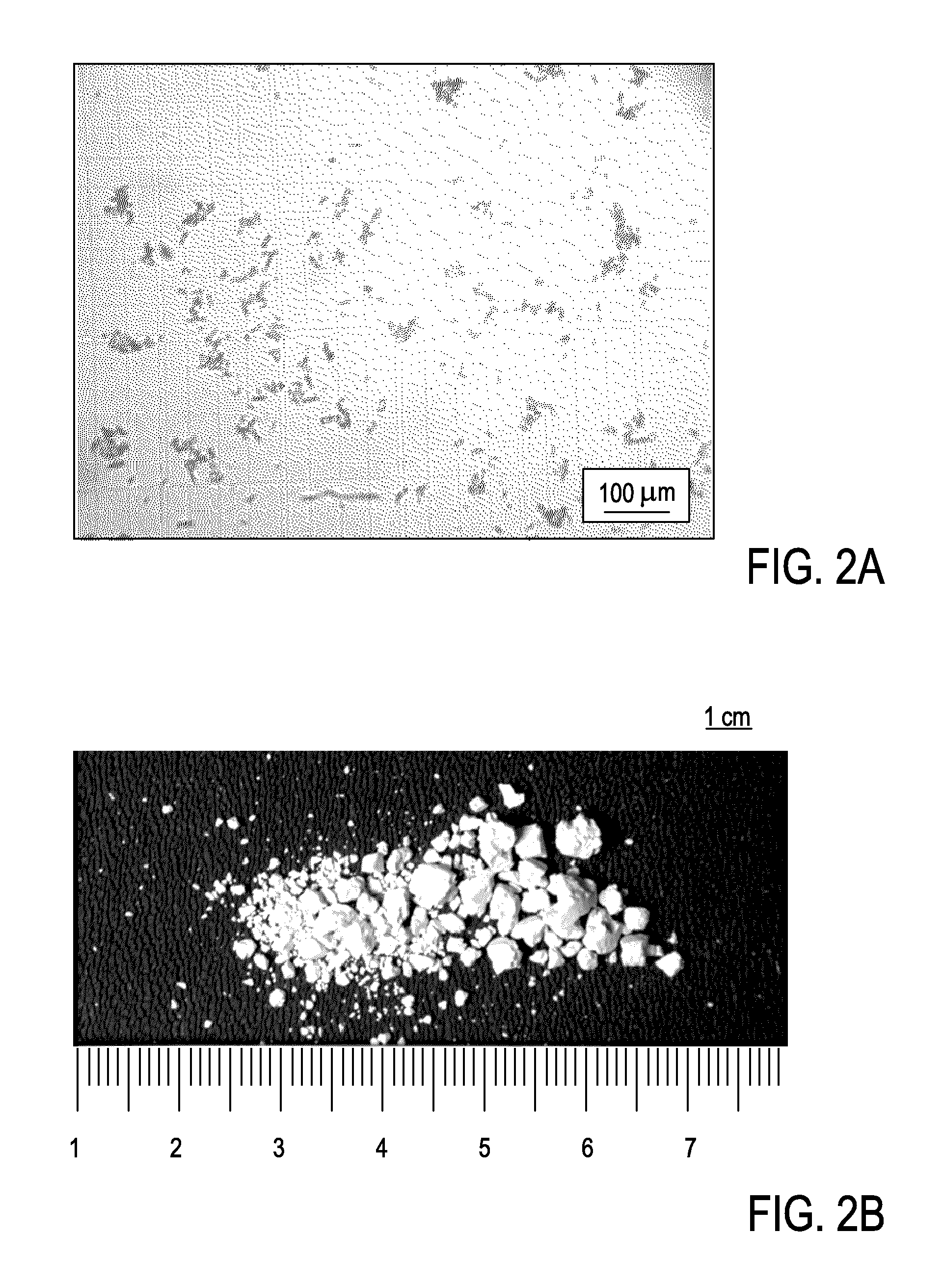
The system effectively breaks down stones into small, smooth fragments for easy passage and reduces collateral tissue damage by using Histotripsy to erode Lithotripsy fragments, enhancing the stone-free rate and minimizing post-treatment discomfort.

Problems solved by technology

Small changes in any of the parameters can result in discontinuation of the ongoing process.
The reduced efficiency is likely due to the increased number of mm size bubbles blocking the shock waves and other energy from reaching the stone.
For this reason, treatment success rates were high without the need for precise image guidance, yet substantial damage to the kidney tissue within the large focal volume also occurred.
By virtue of the smaller focus, the newer generation Lithotripters have reduced collateral tissue damage, but at the expense of success rates.
Although ureteroscopy is an outpatient procedure, patients often suffer with pain and discomfort from a ureteral stent for 4-7 days after treatment.
Disadvantages of Lithotripsy include a stone free rate of ˜65% percent 4 weeks after treatment (compared with 90-95% stone free rate in patients having percutaneous and ureteroscopic procedures) and the necessity and occasionally discomfort of passing stone fragments following treatment.
Furthermore, urinary stones fragmented using shockwave Lithotripsy can remain up to several mm in size and include sharp or jagged edges that make them difficult and painful to pass through the urinary tract.

Method used


Image

  • Devices and Methods for Using Controlled Bubble Cloud Cavitation in Fractionating Urinary Stones
  • Devices and Methods for Using Controlled Bubble Cloud Cavitation in Fractionating Urinary Stones
  • Devices and Methods for Using Controlled Bubble Cloud Cavitation in Fractionating Urinary Stones

Examples


Embodiment Construction

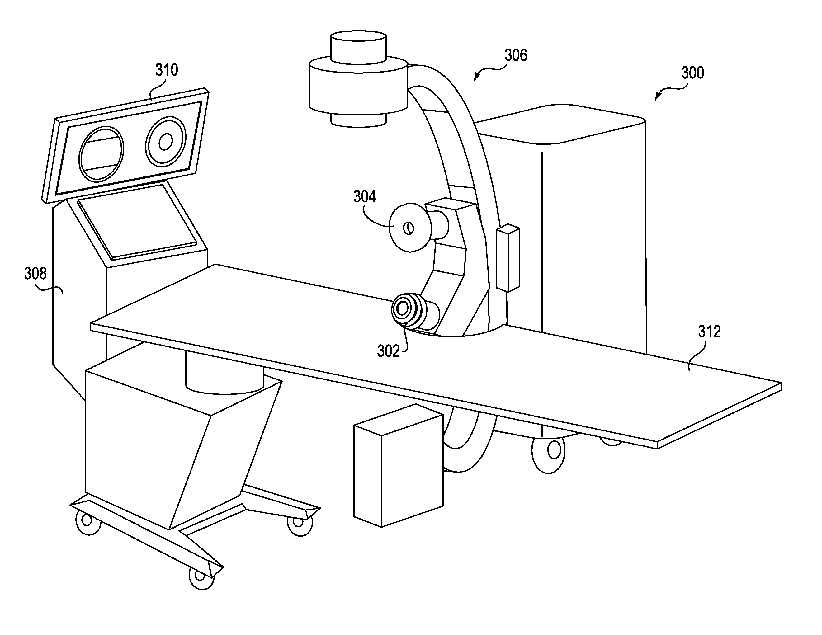
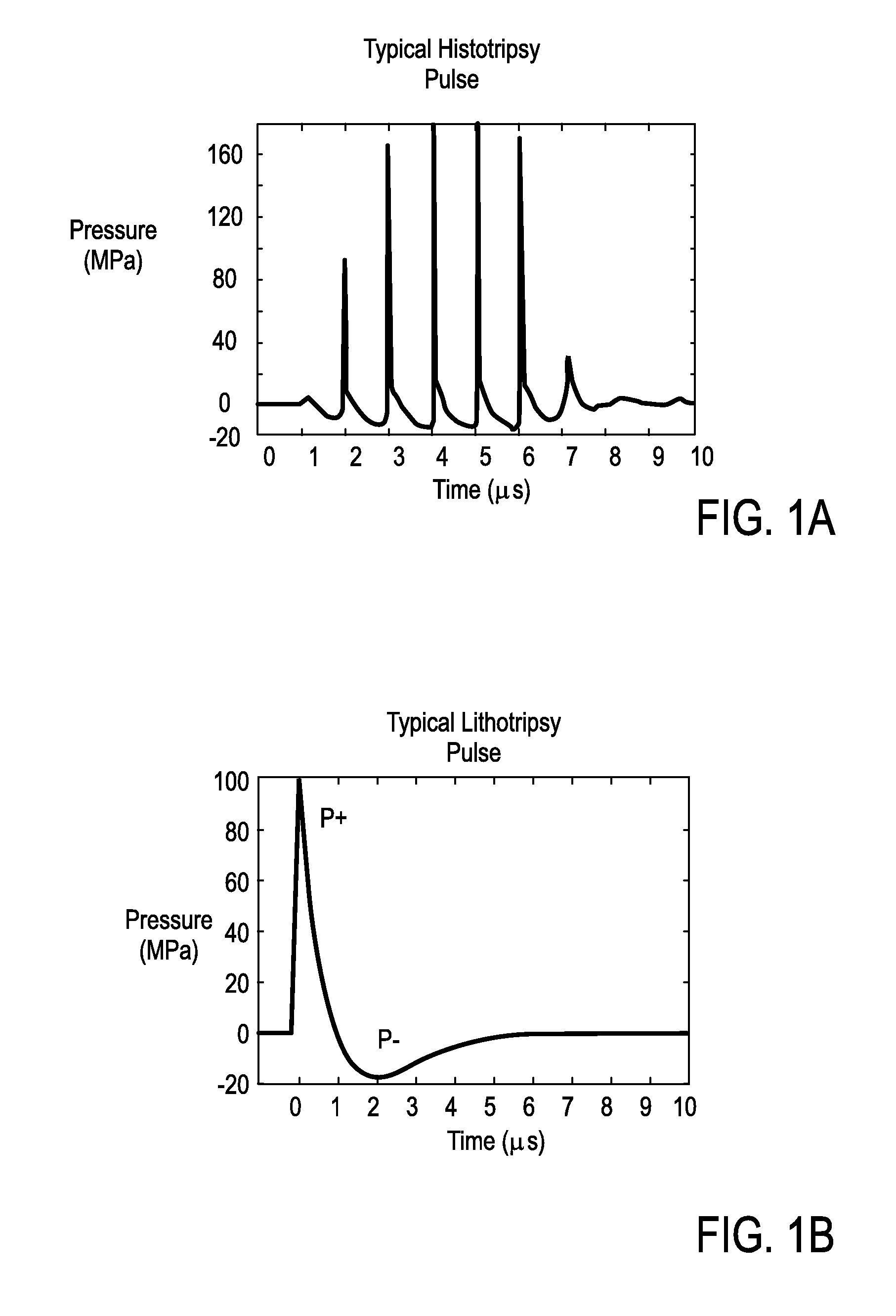
[0034]In addition to imaging tissue, ultrasound technology is increasingly being used to treat and destroy tissue. In medical applications such as Histotripsy, ultrasound pulses are used to form cavitational microbubbles in tissue to mechanically break down and destroy tissue. In Lithotripsy procedures, ultrasound pulses are used to form acoustic shockwaves that break up urinary stones into smaller fragments. Particular challenges arise in using Lithotripsy to break up urinary stones, including failing to break stones down into sizes small and smooth enough to pass comfortably, as well as visualizing and tracking the stones within the patient. The present invention describes several embodiments of devices and methods for treating urinary stones or other calculi including, but not limited to biliary calculi such as gall stones, particularly through the combination of Histotripsy and Lithotripsy therapy in a single procedure.

[0035]Despite the difference between Lithotripsy and Histotr...


PUM


Abstract

A medical imaging and therapy device is provided that may include any of a number of features. One feature of the device is that it can deliver Lithotripsy therapy to a patient, so as to fractionate urinary stones. Another feature of the device is that it can deliver Histotripsy therapy to a patient, so as to erode urinary stones. In some embodiments, the medical imaging and therapy device is configured to target and track urinary stones in the patient during therapy. Methods associated with use of the medical imaging and therapy device are also covered.

Description

CROSS REFERENCE TO RELATED APPLICATIONS[0001]This application is a continuation of pending U.S. patent application Ser. No. 12 / 868,775, filed Aug. 26, 2010, which application claims the benefit under 35 U.S.C. 119 of U.S. Provisional Patent Application No. 61 / 237,011, filed Aug. 26, 2009, titled “Devices and Methods for Using Controlled Bubble Cloud Cavitation in Fractionating Kidney Stones”. These applications are herein incorporated by reference in their entirety.INCORPORATION BY REFERENCE[0002]All publications, including patents and patent applications, mentioned in this specification are herein incorporated by reference in their entirety to the same extent as if each individual publication was specifically and individually indicated to be incorporated by reference.FIELD OF THE INVENTION[0003]The present invention generally relates to ultrasound treatment of urinary stones. More specifically, the present invention relates to using a combination of Lithotripsy and Histotripsy to f...

Claims


Application Information

Patent Timeline
Patent Type & Authority Applications(United States)
IPC IPC(8): A61N7/00A61B8/00
CPCA61N7/00A61B8/00A61B17/225A61B17/2256A61B2017/00172A61B2017/22008A61B2090/378
Inventor CAIN, CHARLES A.HALL, TIMOTHY L.ROBERTS, WILLIAM W.XU, ZHENFOWLKES, J. BRIANDAVISON, THOMAS W.
Owner RGT UNIV OF MICHIGAN
Learn More