Method and system for identification of calcification in imaged blood vessels

a technology of imaged blood vessels and detection methods, applied in image enhancement, angiography, instruments, etc., can solve the problems of difficult identification, difficult to tell calcium voxels apart from lumen voxels, and difficult to accurately segmen

Active Publication Date: 2014-01-02
TOSHIBA MEDICAL SYST CORP
View PDF4 Cites 26 Cited by
  • Summary
  • Abstract
  • Description
  • Claims
  • Application Information

AI Technical Summary

Problems solved by technology

This identification can be difficult in coronary CTA images that are obtained in a contrast CTA scan, in which contrast dye is injected into a vein of the patient, and images are recorded as the dye circulates into the arteries.
Hence it is d...

Method used


Image

  • Method and system for identification of calcification in imaged blood vessels
  • Method and system for identification of calcification in imaged blood vessels
  • Method and system for identification of calcification in imaged blood vessels

Examples


Embodiment Construction

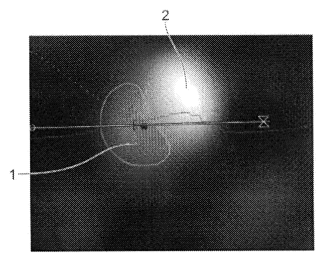
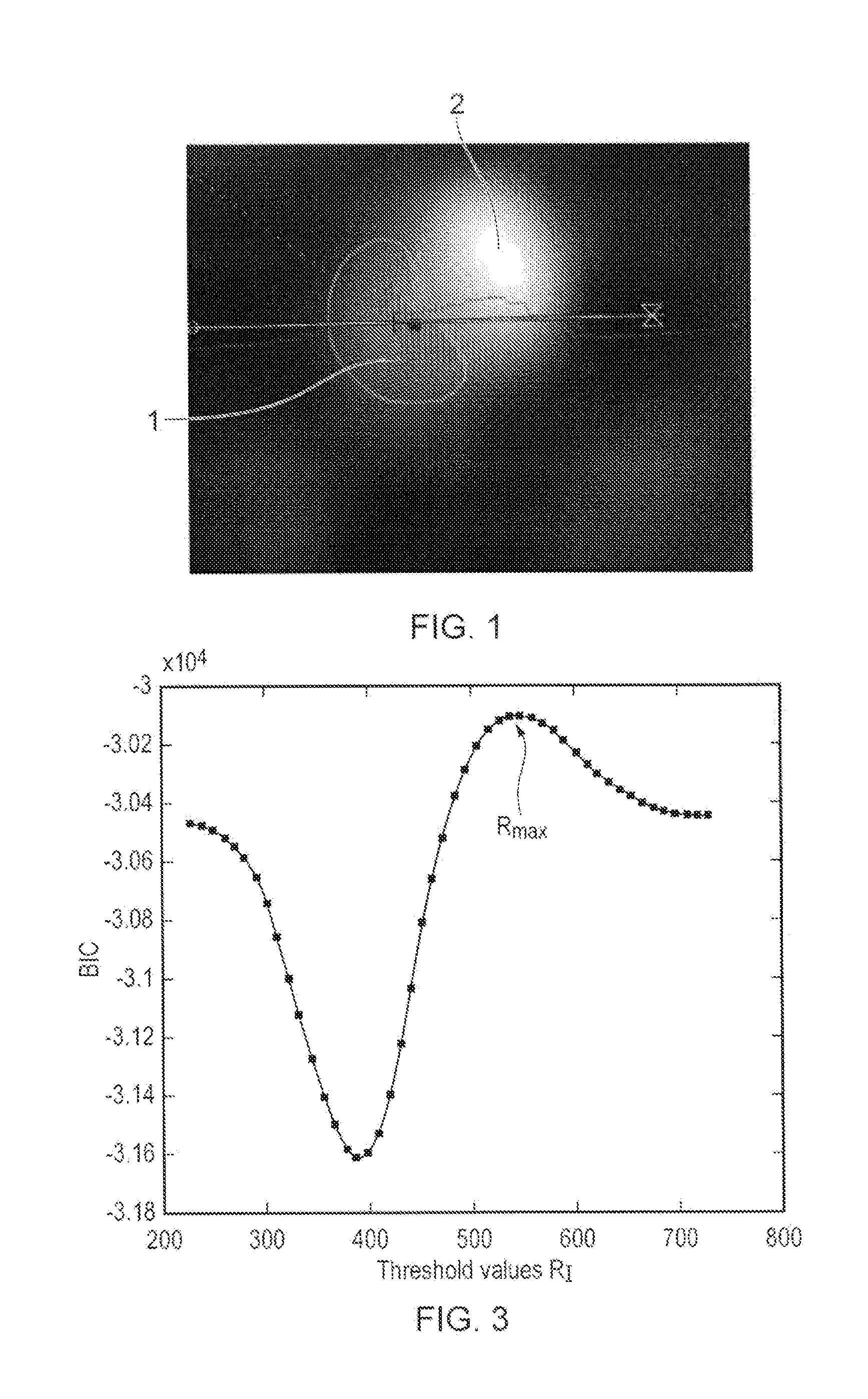
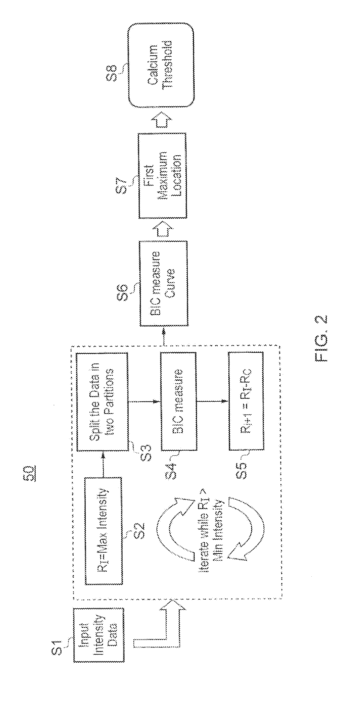
[0022]Certain embodiments of the invention provide a computer system for and method of identifying calcification in a patient image data set showing one or more blood vessels. The computer system is operable to execute machine readable instructions for carrying out a method comprising: obtaining an image data set comprising voxels each having an intensity value; forming the intensity values into a group of intensity values, the intensity values covering an intensity range; defining a plurality of intensity thresholds with values spaced across the intensity range and including the end values of the intensity range; for each of the plurality of intensity thresholds partitioning the group of intensity values into two sub-groups according to the intensity threshold, and calculating an information criterion as a function of the intensity threshold; generating an information criterion measure curve, being a plot of the calculated information criteria against intensity threshold; and locat...


PUM


Abstract

A computer implemented method identifying calcification in a patient image data set including blood vessels. The method includes: obtaining an image data set including voxels each having an intensity value; forming the intensity values into an intensity value group covering an intensity range; defining plural intensity thresholds across the intensity range and including its end values; for each intensity threshold, partitioning the intensity values into two sub-groups according to intensity threshold, and calculating an information criterion based on intensity threshold; generating an information criterion measure curve that plots the calculated information criteria against intensity threshold; locating a maximum in the information criterion measure curve and setting the corresponding intensity threshold as a calcification threshold; and partitioning the intensity values into two sub-groups using the calcification threshold, identifying voxels corresponding to intensity values in the sub-group above the calcification threshold as representing calcification in the patient.

Description

BACKGROUND OF THE INVENTION[0001]The present invention relates to the detection of calcification and the segmentation of vessel lumen and vessel walls in three-dimensional volume data sets obtained by medical imaging procedures such as computed tomography (CT).[0002]CT scanning and other three-dimensional patient imaging techniques such as magnetic resonance imaging yield large image data sets comprised of three-dimensional arrays of voxels. Each voxel has a value indicative of the tissue type present at the corresponding location in the patient's body. In a CT data set the voxels have values defined in Hounsfield units (HU), representing the density of the tissue to the X-rays used to record the image.[0003]When analysing the data set for research or diagnostic purposes, it is necessary to identify which voxels correspond to which types of tissue, so the voxels can be classified according to tissue type. Different organs and tissues can then be distinguished in the image. Subsequen...

Claims


Application Information

Patent Timeline
IPC IPC(8): G06K9/34G06V10/28
CPCG06T7/0012G06T2207/10081G06T2207/30101A61B6/032A61B6/481A61B6/504A61B6/5217G16H50/30G06V20/653G06V10/28G06V10/421G06V10/7715G06F18/2134
Inventor MASOOD, SAADMOHR, BRIANPLAKAS, COSTAS
Owner TOSHIBA MEDICAL SYST CORP
Why Eureka
Try Eureka