Structural Model Of G Protein-Coupled Receptor And Method For Designing Ligand Capable Of Binding To G Protein-Coupled Receptor Using The Structural Model

a structure model and g protein-coupled receptor technology, applied in the direction of peptides, instruments, molecular structures, etc., can solve the problems of rhodopsin not solely providing a structural model, the role of the kink in the functioning of gpcrs remains unclear, and the nature of specific conformational change has yet to be understood

Inactive Publication Date: 2014-01-23
SUNTORY HLDG LTD
View PDF0 Cites 1 Cited by
  • Summary
  • Abstract
  • Description
  • Claims
  • Application Information

AI Technical Summary

Problems solved by technology

However, the role of the kink in the functioning of GPCRs still remains unclear.
For this reason, the crystal structure of rhodopsin does not solely provide a structural model for every receptor / ligand complex.
Nonetheless, the nature of specific conformational change has yet to be understood.

Method used


Image

  • Structural Model Of G Protein-Coupled Receptor And Method For Designing Ligand Capable Of Binding To G Protein-Coupled Receptor Using The Structural Model
  • Structural Model Of G Protein-Coupled Receptor And Method For Designing Ligand Capable Of Binding To G Protein-Coupled Receptor Using The Structural Model
  • Structural Model Of G Protein-Coupled Receptor And Method For Designing Ligand Capable Of Binding To G Protein-Coupled Receptor Using The Structural Model

Examples


example 1

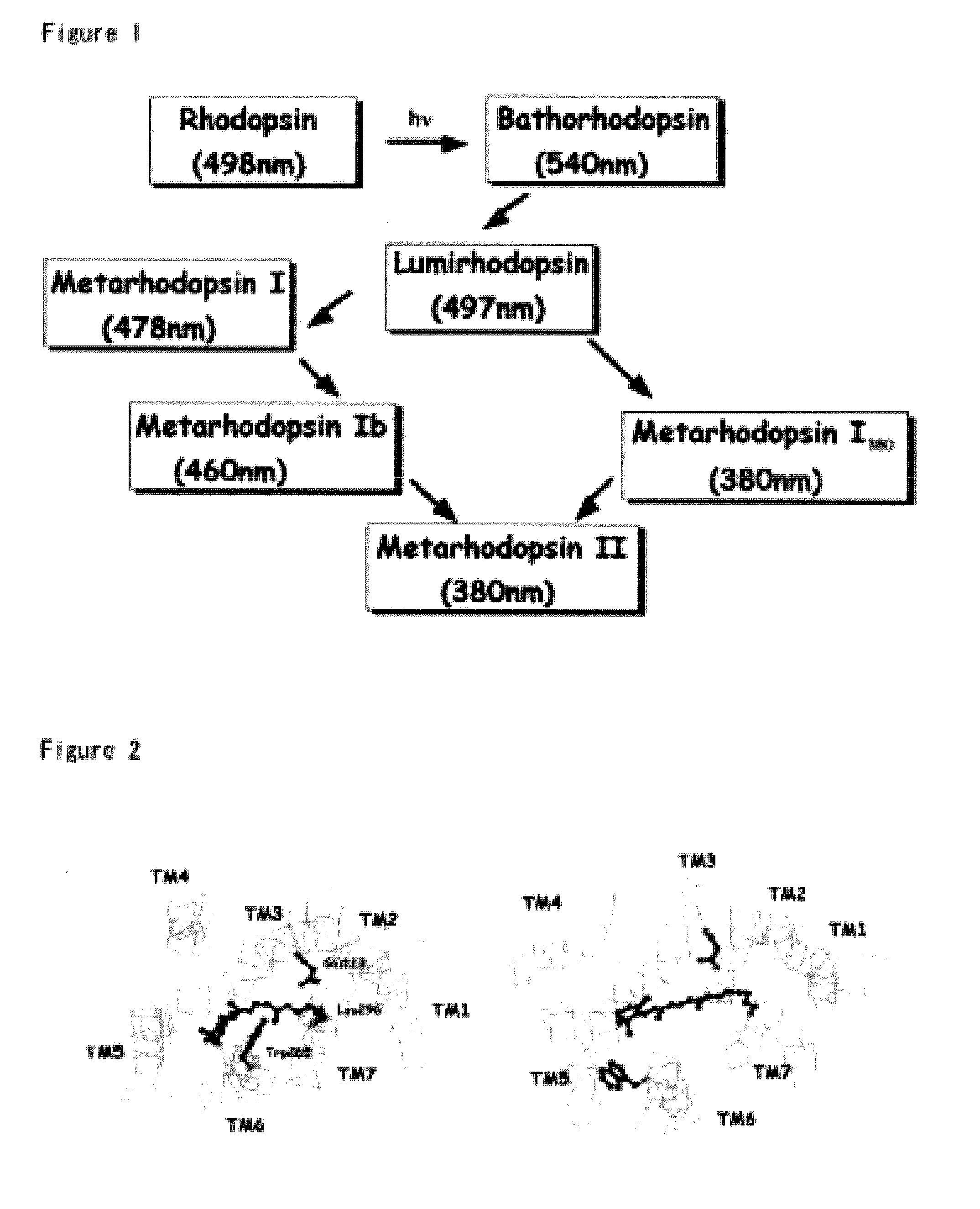
Construction of Models for Photoactivated Intermediates of Rhodopsin

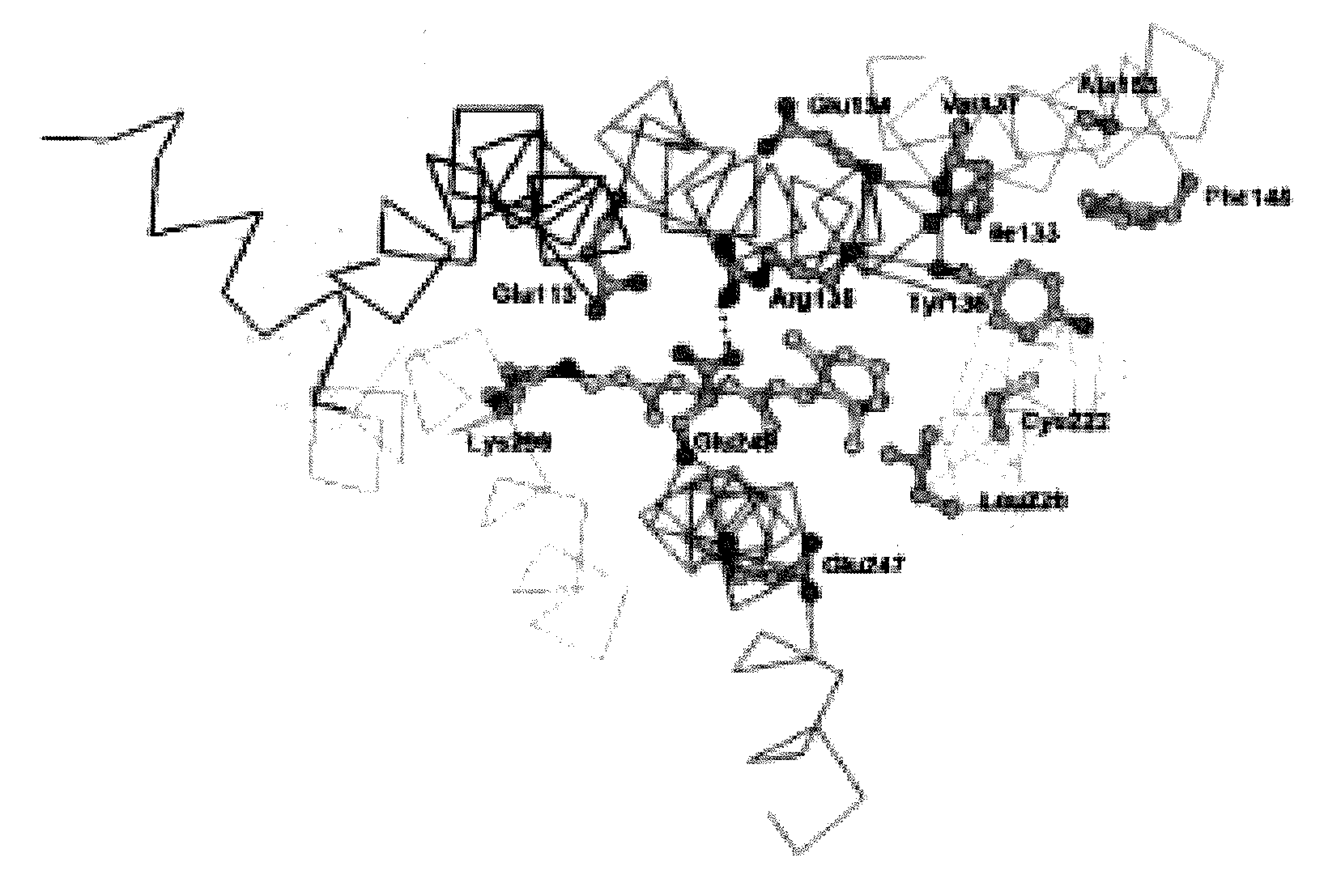
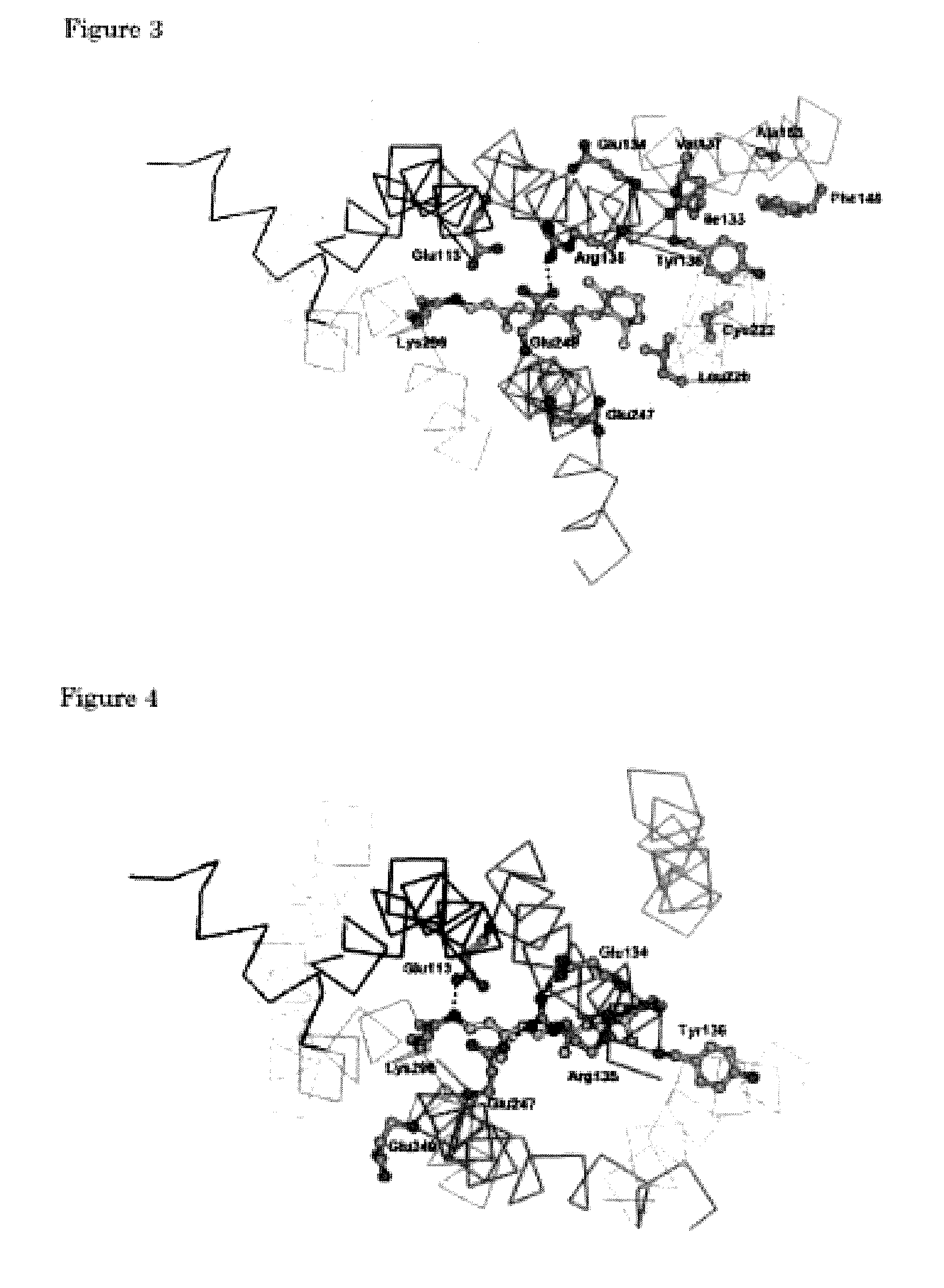
[0129]Using a molecule modeling software Insight II-Discover 3 (Molecular Simulations Inc., USA), a structural model for each of the rhodopsin intermediates was generated and was optimized based on the crystal structure of rhodopsin (Palczewski et al., Science, 289, 144-167, 2000). TM3 was swung about the Cα carbon of Cys110 to serve as the pivot point while the distance to TM2 was kept at 5 Å or more. The magnitude of the swing was determined by taking into consideration the interaction of TM6 with Glu247 for each of Lumi, Meta I, Meta Ib, and Meta I380 structures. Specifically, in each of Lumi, Meta I, Meta Ib, and Meta I380, Cys140 on TM3 was swung in such a manner that Cys140 is spaced from TM6 by a distance of 1.6 Å, 4.3 Å, 6.8 Å, and 9.0 Å, respectively. Furthermore, N-terminal (Glu150) of the portion of TM4 that would interfere with TM3 was swung toward TM5 about Gly174 on the C-terminal of the helix to serv...

example 2

Construction of Models for GPCR and GPCR / Ligand Complex

[0132]Using the structure of Meta I, Meta Ib, Meta I380, and Meta II and based on the homology among the amino acid sequences of rhodopsin and other GPCRs (FIG. 18), three-dimensional conformations for binding a full agonist, a partial agonist, an antagonist, and an inverse agonist were constructed for each of the GPCRs.

[0133]For each of the GPCRs, a receptor conformation for binding an inverse agonist was generated by using the structure of Meta I as a template. Using a homology module of Insight II, amino acid substitution was carried out, as were insertion or deletion of amino acid residues in the loop region. Using Discover 3, the conformation was optimized so that the Cα carbon of the amino acids was fixed as firmly as possible.

[0134]Likewise, three-dimensional conformations for binding an antagonist, a partial agonist, and a full agonist that correspond to Meta Ib, Meta I380, and Meta II, respectively, were constructed for...

example 3

Construction of Structural Models for Adrenaline Receptors Bound to Antagonist

[0136]Using the structure of rhodopsin Meta Ib as a template, Meta Ib-like structural models of antagonist-bound receptor was constructed for a panel of twelve adrenaline receptors, which form a class of G protein-coupled receptors (GPCRs).

[0137]To construct the structural models for the panel of adrenaline receptors, the amino acid sequence of rhodopsin to serve as a template was first aligned with the amino acid sequences of the panel of adrenaline receptors for which to construct the structural model. Clustal W was used as the alignment program (Thompson et al., Nucleic Acids Research, 22:4673-4680(1994)). The analysis revealed that while the amino acid sequences showed a relatively low homology to one another, the transmembrane regions, which include conserved hydrophobic residues and sequence motifs, are aligned at a relatively high homology, and the less conserved loop regions tend to include abnorma...


PUM

PropertyMeasurementUnit
angleaaaaaaaaaa
wavelength rangeaaaaaaaaaa
crystal structure modelaaaaaaaaaa

Abstract

The present invention provides a method for constructing a three-dimensional structural model of an activated intermediate of a G protein-coupled protein receptor (GPCR) or a complex between a GPCR and a ligand. The three-dimensional structural model may be used to identify, screen, search, evaluate, or design GPCR agonists or antagonists. In a representative embodiment, a three-dimensional structural model of a photoactivated intermediate of rhodopsin is constructed using molecule modeling software and structural coordinates of the crystal structure of rhodopsin. The three-dimensional structural model of rhodopsin is subsequently used to construct structural models of activated intermediates of other GPCRs.

Description

TECHNICAL FIELD[0001]The present invention relates to a structural model for receptor / ligand complexes of G protein-coupled receptors (which may be referred to simply as ‘GPCRs,’ hereinafter) and ligands capable of binding to G protein-coupled receptors. It also relates to a method for creating a three-dimensional structural model for activated intermediates of G protein-coupled receptors in the structural model for the receptor / ligand complexes, as well as to structural models of the complexes or the activated intermediates of G protein-coupled receptors obtained by this method. The present invention further relates to a three-dimensional coordinate for determining these structural models.[0002]The present invention further relates to a method for using the three-dimensional structural model of G protein-coupled receptors or a method for using the three-dimensional coordinates for determining the structural model in identifying, screening for, searching for, evaluating, and design...

Claims


Application Information

Patent Timeline
Patent Type & Authority Applications(United States)
IPC IPC(8): G06F19/12G16B5/00C07K14/72G01N33/566G01N33/68G16B15/30
CPCG06F19/12C07K14/723C07K2299/00G01N33/566G01N33/6803G16B15/00G16B15/30G16B5/00
Inventor ISHIGURO, MASAJI
Owner SUNTORY HLDG LTD
Why Eureka
Try Eureka