Quantifying curvature of biological structures from imaging data

Active Publication Date: 2014-03-06
AGENCY FOR SCI TECH & RES +1
View PDF5 Cites 6 Cited by
  • Summary
  • Abstract
  • Description
  • Claims
  • Application Information

AI Technical Summary

Benefits of technology

The patent is about a method for analyzing the shape and performance of the heart using medical imaging techniques. The method allows for the accurate and consistent measurement of curvedness and its rate of change, which can provide valuable information to physicians and researchers studying heart disease. The method can also be used to assess the suitability of cardiac resynchronization therapy in patients with heart disease. Overall, the patent presents a useful tool for studying and improving heart performance.

Problems solved by technology

Adverse ventricular remodeling leads to development of heart failure (HF).
These methods lack specificity and only describe limited aspects of the LV.
Nevertheless, there is still a lack of an efficient and clinically meaningful way to capture and quantify complex 3D LV conformational data.

Method used


Image

  • Quantifying curvature of biological structures from imaging data
  • Quantifying curvature of biological structures from imaging data
  • Quantifying curvature of biological structures from imaging data

Examples


Embodiment Construction

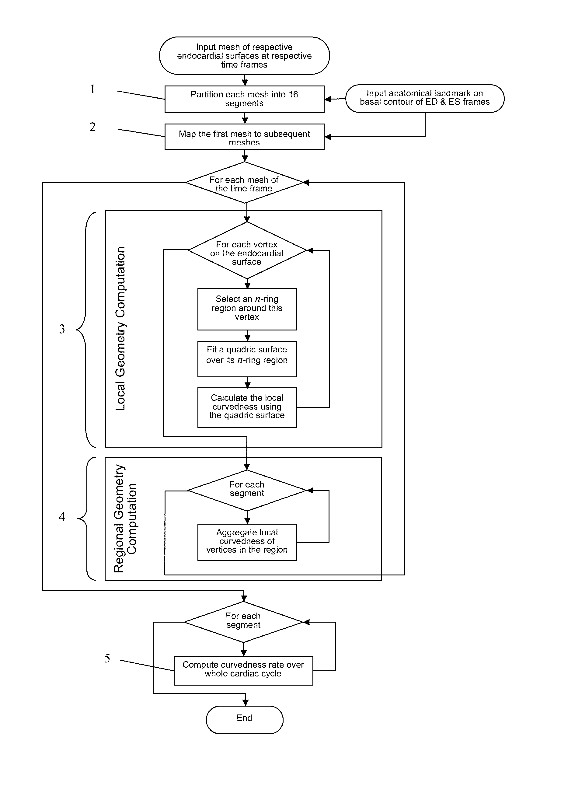
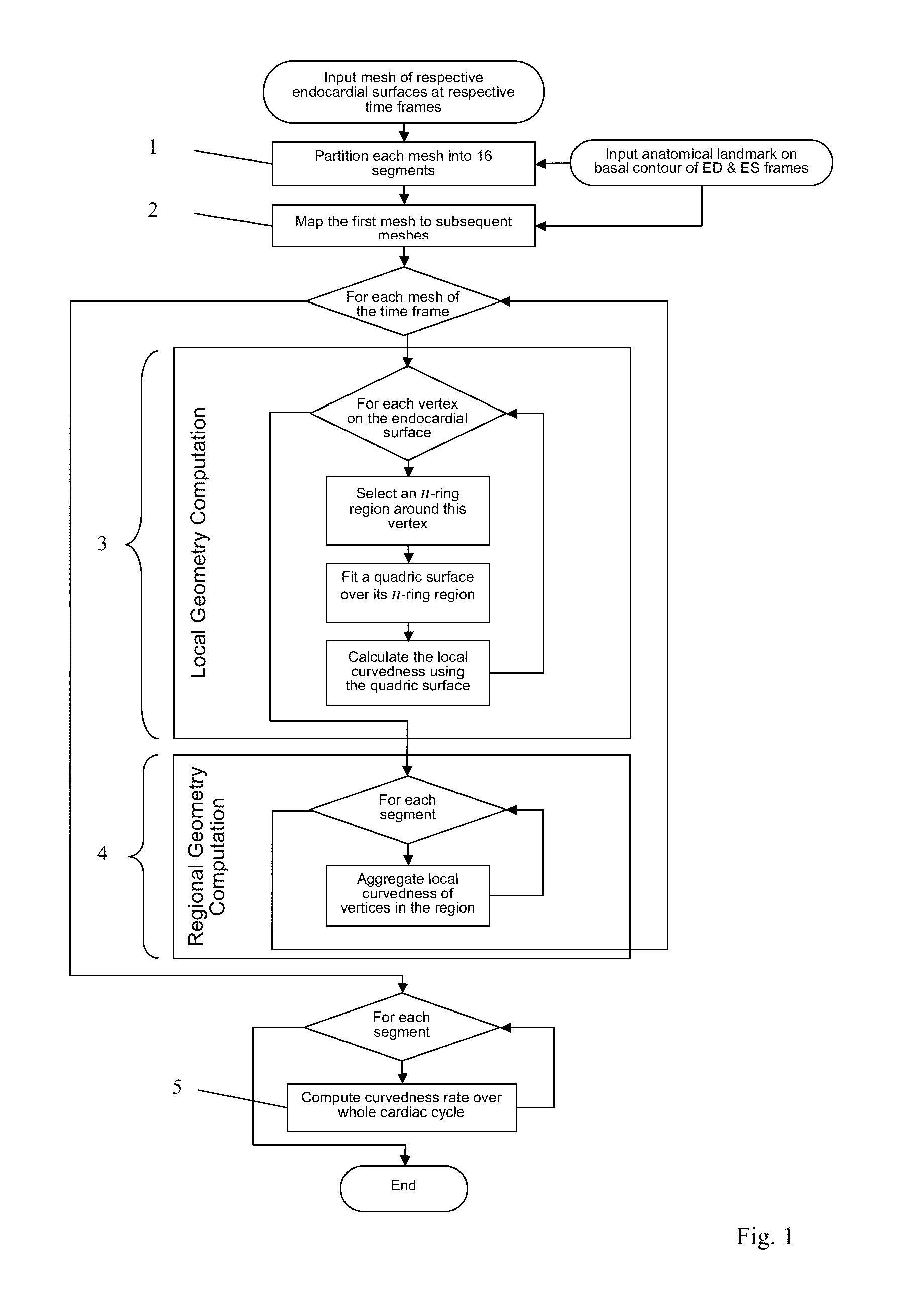
[0034]Referring firstly to FIG. 1, a method which is an embodiment of the invention is illustrated as a flow diagram. The method may be performed by a computer system, such as a standard generally programmed computer, having a data storage device storing program instructions to implement the method steps. The workflow performs very fast (in human terms, substantially instantaneous) shape quantitation.

[0035]There are two inputs to the system:[0036](i) a set of surface meshes representing the instantaneous shape of the endocardial surface of the left ventricle (LV) at each of a set of respective instants (“time frames”). These surface meshes are derived from MRI data captured at the respective times. There are many existing ways of generating the input meshes. To illustrate, FIG. 4(a) shows the MRI data for a given instant. FIG. 4(b) shows delineated contours produced from the MRI data. FIG. 4(c) shows the mesh for the corresponding instant, which is the input to the embodiment.[0037]...


PUM


Abstract

A method and system are proposed to obtain quantitative data about the shape of a biological structure, and especially a heart ventricle. A set of three-dimensional input meshes are generated from MRI data. They represent the shape of a ventricle at successive times. The input meshes are used to generate a set of three-dimensional morphed meshes which have the same number of vertices as each other, and have respective shapes which are the shapes of corresponding ones of the input meshes. Then, for each of the times, shape analysis is performed to obtain a curvedness value at each of a plurality of corresponding locations in the morphed meshes. The curvedness value may be used to obtain a curvedness rate at each of the locations, indicative of the rate of change of curvedness with time at each of the locations.

Description

FIELD OF THE INVENTION[0001]The present invention relates to computer systems, and computerized methods, for using image data such as Magnetic Resonance Imaging (MRI) data, to measure changes in the curvature of a biological structure, such as a heart ventricle, and in particular the left ventricle (LV) of a subject's heart.BACKGROUND OF THE INVENTION[0002]Ventricular remodeling is defined as changes in size, shape and function of the heart after cardiac injury (i.e., acute myocardial infarction). Adverse ventricular remodeling leads to development of heart failure (HF). Accurate quantitative assessment of ventricular remodeling (i.e., shape and shape deformation) is instrumental both for diagnosis and prognosis and to follow the effectiveness of therapeutic interventions.[0003]Conventionally, many techniques used in actual clinical settings are based on two-dimensional (2D) left ventricle (LV) contours, such as sphericity index and subsequently conicity index. These methods lack sp...

Claims


Application Information

Patent Timeline
IPC IPC(8): G06T7/00
CPCG06T7/0012G06T7/0014G06T2200/04G06T2207/10088G06T2207/30048G06T7/64
Inventor SU, YILIM, CHI WAN CALVINTAN, RU SANZHONG, LIANG
Owner AGENCY FOR SCI TECH & RES
Why Eureka
Try Eureka