Eureka AIR delivers breakthrough ideas for toughest innovation challenges, trusted by R&D personnel around the world.

Method for automatically adjusting a focal plane of a digital pathology image

a digital pathology image and automatic adjustment technology, applied in image enhancement, image analysis, instruments, etc., can solve the problems of not being perfectly aligned with the focal plane, cells will not be positioned in a perfectly flat layer, and the tissue is not perfectly flat and typically not perfectly aligned, so as to reduce the computational load of the algorithm

Active Publication Date: 2014-06-19
KONINKLJIJKE PHILIPS NV
View PDF20 Cites 2 Cited by
  • Summary
  • Abstract
  • Description
  • Claims
  • Application Information

AI Technical Summary

Benefits of technology

The present invention solves a problem in microscopy where the traditional method of adjusting the focal plane was time-consuming and resulted in poor image quality. The new method uses individual biological objects, such as cells or nuclei, as the basis for decision-making and adjusts the focal plane accordingly to ensure optimal sharpness. The method also reduces the computational load and allows for faster focusing. Additionally, the invention includes an embodiment that assigns a respective focal plane to each pixel location in the image and stores it for easy reference during adjustment. The technical effects of this invention include improved image quality, faster focusing, and improved efficiency of microscopy analysis.

Problems solved by technology

This tissue is not perfectly flat and typically not perfectly aligned with the focal plane of the imaging system.
Typically, these cells will not be positioned in a perfectly flat layer and the layer will not be perfectly aligned with the focal plane of the imaging system.

Method used


Image

  • Method for automatically adjusting a focal plane of a digital pathology image
  • Method for automatically adjusting a focal plane of a digital pathology image
  • Method for automatically adjusting a focal plane of a digital pathology image

Examples


Embodiment Construction

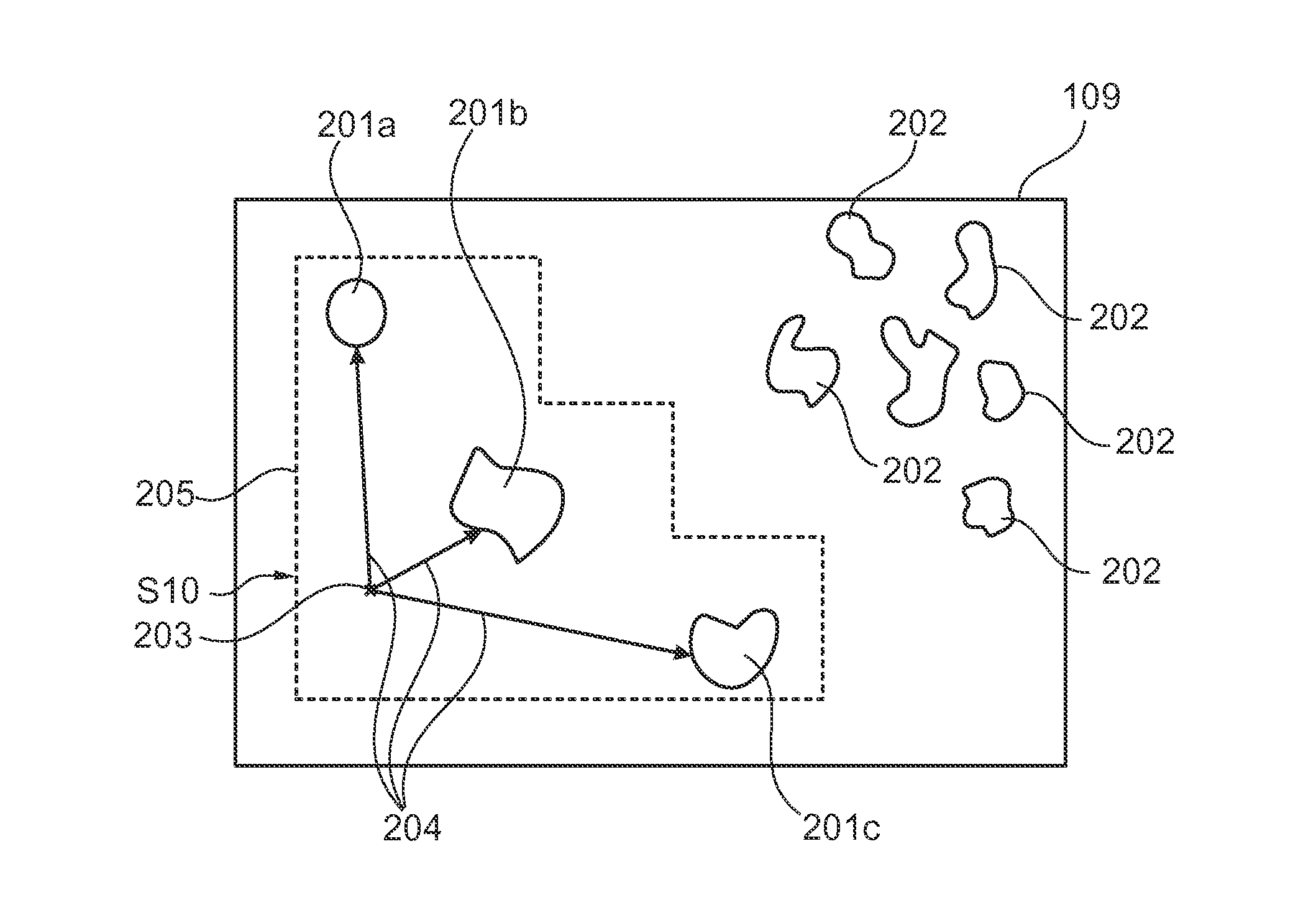
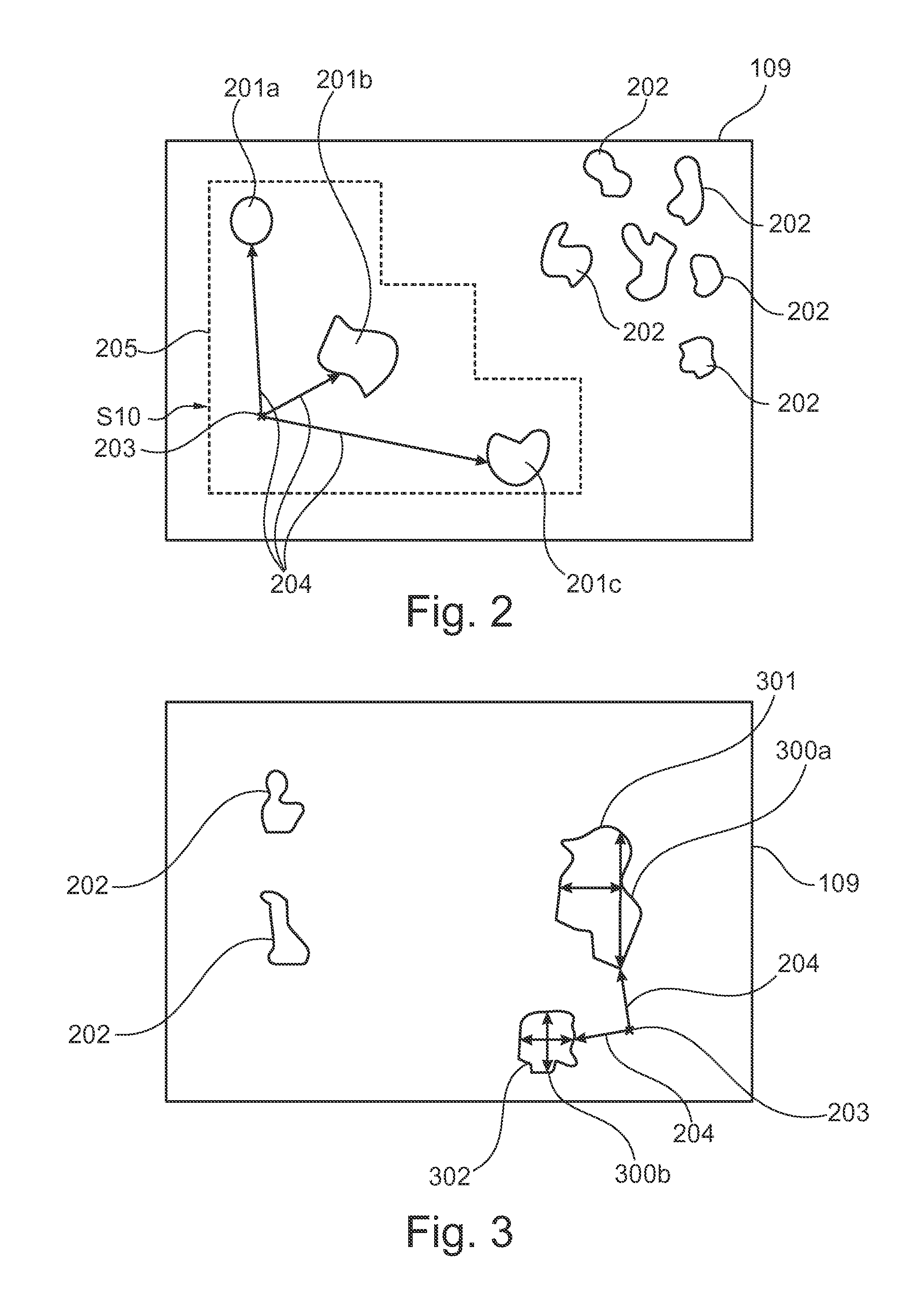
[0068]FIG. 1 shows a flow diagram of a method for automatically adjusting a focal plane of a digital pathology image according to an exemplary embodiment of the present invention. The image used in said method comprises a plurality of individual biological objects. The depicted method comprises the step of providing for three-dimensional digital pathology image data comprising a plurality of focal planes S1, and as a second step displaying the digital pathology image data as an image S2. Thereby, the displayed image has a focal plane which is from the plurality of focal planes. As a further step, determining a position of interest in the image a user has (S3) is depicted as step S3. The step S4 describes the step of calculating which individual biological object out of the plurality of objects is the object of interest. This calculation may also be embodied as deciding which object of the plurality of objects is the closest to the determined position of interest. For example, the ob...


PUM


Abstract

The present invention provides for an intelligent autofocus method and device for a digital pathology image, method are device are able to couple a feedback from the behaviour of the user, for example by gaze tracking arrangement, to the actually focal representation of an image and adjust the imaging setup accordingly with regard to the focal plane. The adjustment is performed in such a way that the focal plane is selected which optimizes and sharpens the actual object of interest which was previously determined.

Description

FIELD OF THE INVENTION[0001]The present invention relates to autofocus technology. In detail, the present invention relates to a method for automatically adjusting a focal plane of a digital pathology image, to a device for displaying images and for automatic focal plane adjustment of a digital pathology image, to a program element as well as to a computer-readable medium.BACKGROUND OF THE INVENTION[0002]In pathology, images of tissue samples or cell smears are analyzed under a microscope. In histopathology, the pathologist studies tissue in order to come to a diagnosis. A tissue sample is a thin slice of tissue mounted between two glass slides. This tissue is not perfectly flat and typically not perfectly aligned with the focal plane of the imaging system. Besides that, the cells of the tissue are positioned at different heights in the tissue layer. Consequently, a significant part of the cells in a two-dimensional tissue image can be out of focus. The same goes for cytopathology i...

Claims


Application Information

Patent Timeline
Patent Type & Authority Applications(United States)
IPC IPC(8): G06T7/00
CPCG06T7/0012G02B21/16G02B21/244G06T5/50G06T2207/10056G06T2207/10148G06T2207/20092G06T2207/30024
Inventor VAN LEEUWEN, MARINUS BASTIAANVINK, JELTA PETER
Owner KONINKLJIJKE PHILIPS NV
Why Eureka
Eureka Blog
Learn More