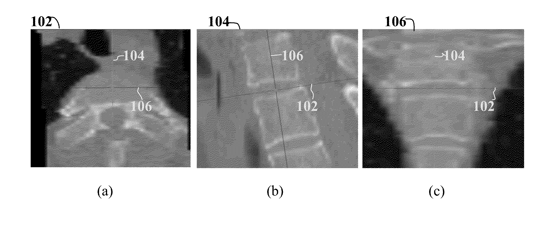
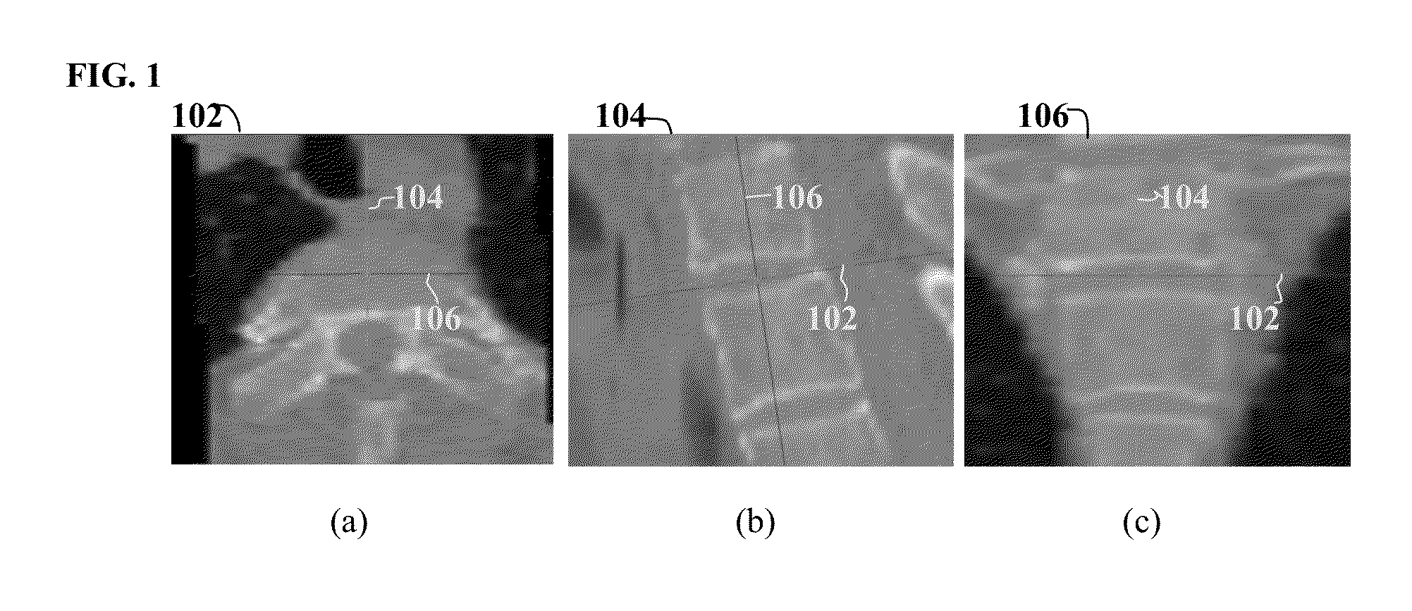
Method and System for On-Site Learning of Landmark Detection Models for End User-Specific Diagnostic Medical Image Reading

a landmark detection and diagnostic imaging technology, applied in the field of landmark detection models, can solve the problems of greater challenges in the field of automatic landmark detection and image orientation

Active Publication Date: 2014-08-07
SIEMENS HEALTHCARE GMBH
View PDF19 Cites 45 Cited by
  • Summary
  • Abstract
  • Description
  • Claims
  • Application Information

AI Technical Summary

Benefits of technology

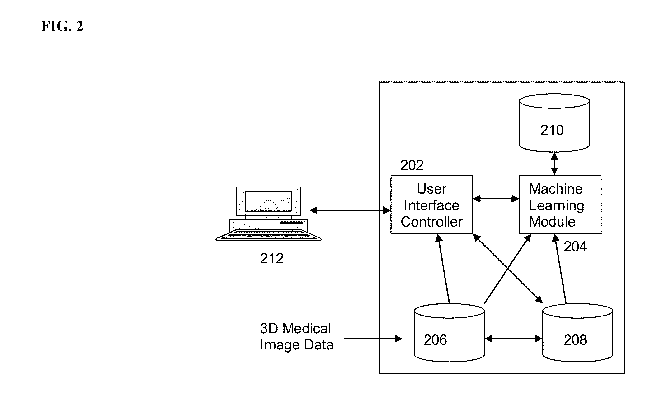
[0004]The present invention provides a method and system for automatic on-site learning of landmark detection models for end user-specific diagnostic medical image reading. Embodiments of the present invention address the problem of fully automatic 3D landmark detection, and provide a workflow individualization methodology that can cope with a dynamic radiological environment. Embodiments of the present invention allow an end user to vary scenarios of radiological image reading in terms of relevant landmarks or view planes and associated medical imaging data of the appropriate modality.

Problems solved by technology

However, manual navigation to particular anatomical landmarks and double oblique alignment of 2D planes to the anatomical landmarks is time consuming and the radiologists' workflow can significantly be improved by automatic detection of landmarks and corresponding oriented planes.
Furthermore, hospitals and groups of physicians all over the world may have their own individual preferences, leading to greater challenges in automating landmark detecting and image orientation.

Method used


Image

  • Method and System for On-Site Learning of Landmark Detection Models for End User-Specific Diagnostic Medical Image Reading
  • Method and System for On-Site Learning of Landmark Detection Models for End User-Specific Diagnostic Medical Image Reading
  • Method and System for On-Site Learning of Landmark Detection Models for End User-Specific Diagnostic Medical Image Reading

Examples


Embodiment Construction

[0013]The present invention is directed to a method and system for automatic on-site learning of landmark detection models for end user-specific diagnostic medical image reading. Embodiments of the present invention are described herein to give a visual understanding of the methods described herein. A digital image is often composed of digital representations of one or more objects (or shapes). The digital representation of an object is often described herein in terms of identifying and manipulating the objects. Such manipulations are virtual manipulations accomplished in the memory or other circuitry / hardware of a computer system. Accordingly, it is to be understood that embodiments of the present invention may be performed within a computer system using data stored within the computer system.

[0014]Embodiments of the present invention address the problem of fully automatic 3D landmark detection, and provide a workflow individualization methodology that can cope with a dynamic radio...


PUM


Abstract

A method and system for on-line learning of landmark detection models for end-user specific diagnostic image reading is disclosed. A selection of a landmark to be detected in a 3D medical image is received. A current landmark detection result for the selected landmark in the 3D medical image is determined by automatically detecting the selected landmark in the 3D medical image using a stored landmark detection model corresponding to the selected landmark or by receiving a manual annotation of the selected landmark in the 3D medical image. The stored landmark detection model corresponding to the selected landmark is then updated based on the current landmark detection result for the selected landmark in the 3D medical image. The landmark selected in the 3D medical image can be a set of landmarks defining a custom view of the 3D medical image.

Description

BACKGROUND OF THE INVENTION[0001]The present invention relates to learning of landmark detection models for landmark detection in medical images, and more particularly, to on-site learning of landmark detection models for end user-specific diagnostic medical image reading.[0002]In radiology, routine reading of three dimensional medical images, e.g., from computed tomography (CT), magnetic resonance imaging (MRI), positron emission tomography (PET), etc., usually follows fixed rules and protocols. Such protocols are sometimes established and re-fined by individual hospitals on their own behalf to ensure and to achieve a high quality of care. Also, physicians often develop their own methodology for a systematic reading of cases. Depending on the medical question to be answered, navigation through the data sets may require the inspection of certain important anatomical landmarks, in terms of 3D coordinates, or their surroundings. Standardized tomographic 2D views, i.e., planes, in the ...

Claims


Application Information

Patent Timeline
Patent Type & Authority Applications(United States)
IPC IPC(8): A61B5/00G06V10/772
CPCA61B5/0033A61B6/025A61B6/502G16H30/40G16H50/50G16H30/20A61B5/0036G06V2201/03G06V10/772G06F18/28G06F18/00A61B6/00
Inventor WELS, MICHAELKELM, MICHAELSUEHLING, MICHAELZHOU, SHAOHUA KEVIN
Owner SIEMENS HEALTHCARE GMBH
Why Eureka
Try Eureka