
System for and method of quantifying on-body palpitation for improved medical diagnosis

a technology of on-body palpitation and quantitative methods, applied in the field of object imaging, can solve the problems of subjective palpitation, sinclair does not disclose any algorithm for accurately rendering microscopic structures and arterial pressure pulses

Inactive Publication Date: 2015-01-08
RGT UNIV OF CALIFORNIA
View PDF2 Cites 18 Cited by
  • Summary
  • Abstract
  • Description
  • Claims
  • Application Information

AI Technical Summary

Benefits of technology

The patent text describes a system that uses a deformable membrane with a reflective surface and a camera to create three-dimensional images of an object. The system can be controlled to illuminate the reflective surface from multiple directions and capture images of the object's shape from the reflected light. The captured images are then processed to create a detailed three-dimensional image of the object. The system can also correlate the three-dimensional images with medical diagnoses, providing a tool for medical diagnosis. Overall, the invention helps to improve the accuracy and efficiency of medical diagnosis.

Problems solved by technology

Unfortunately, palpation is subjective inasmuch as the results may vary among physicians.
The results depend on the physician's ability and experience, all of which make the results prone to error.
Sinclair does not disclose any algorithms for accurately rendering microscopic structures and arterial pressure pulses, nor is it capable of being packaged in a mobile, low-cost package.

Method used


Image

  • System for and method of quantifying on-body palpitation for improved medical diagnosis
  • System for and method of quantifying on-body palpitation for improved medical diagnosis
  • System for and method of quantifying on-body palpitation for improved medical diagnosis

Examples


Embodiment Construction

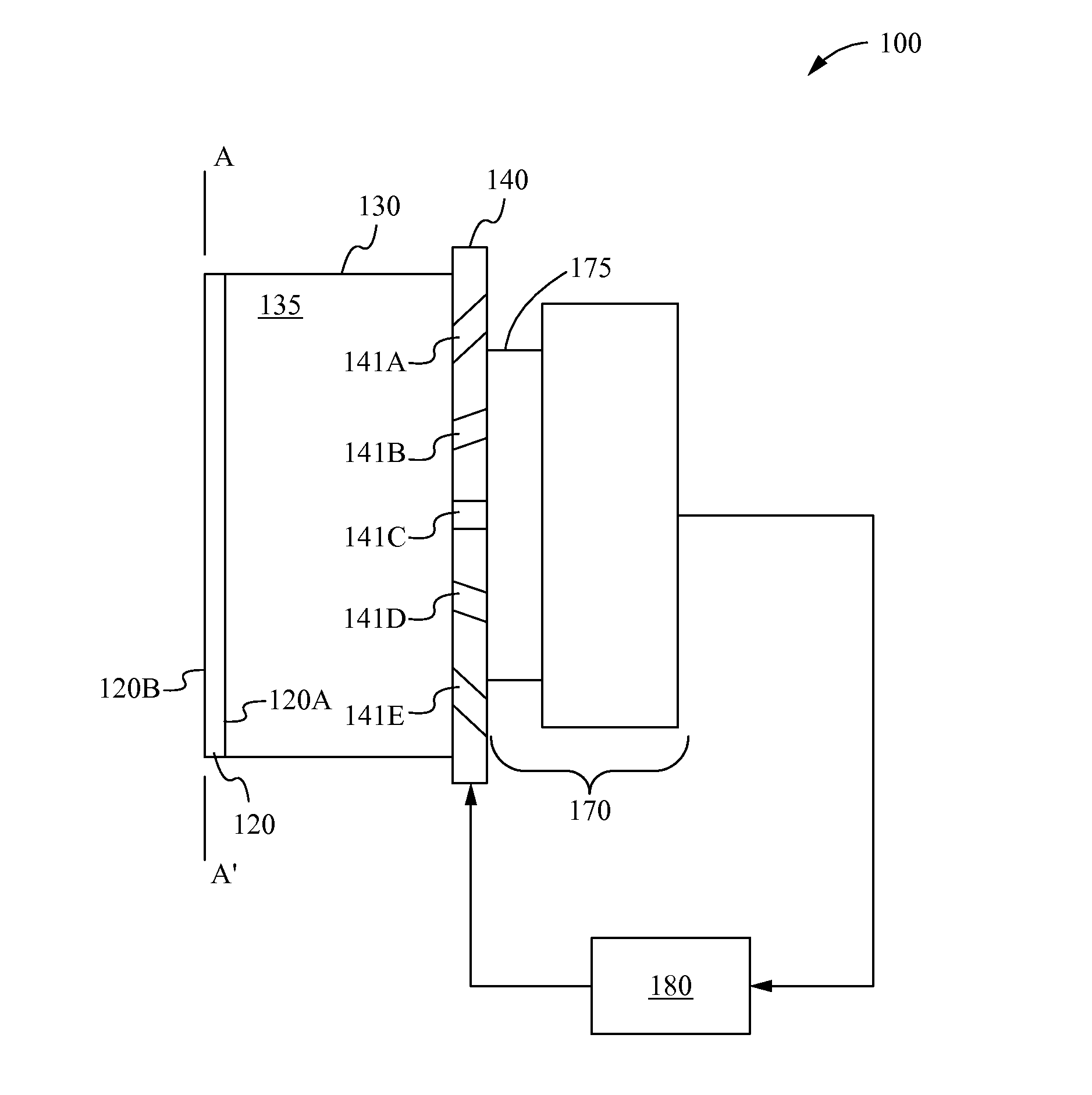
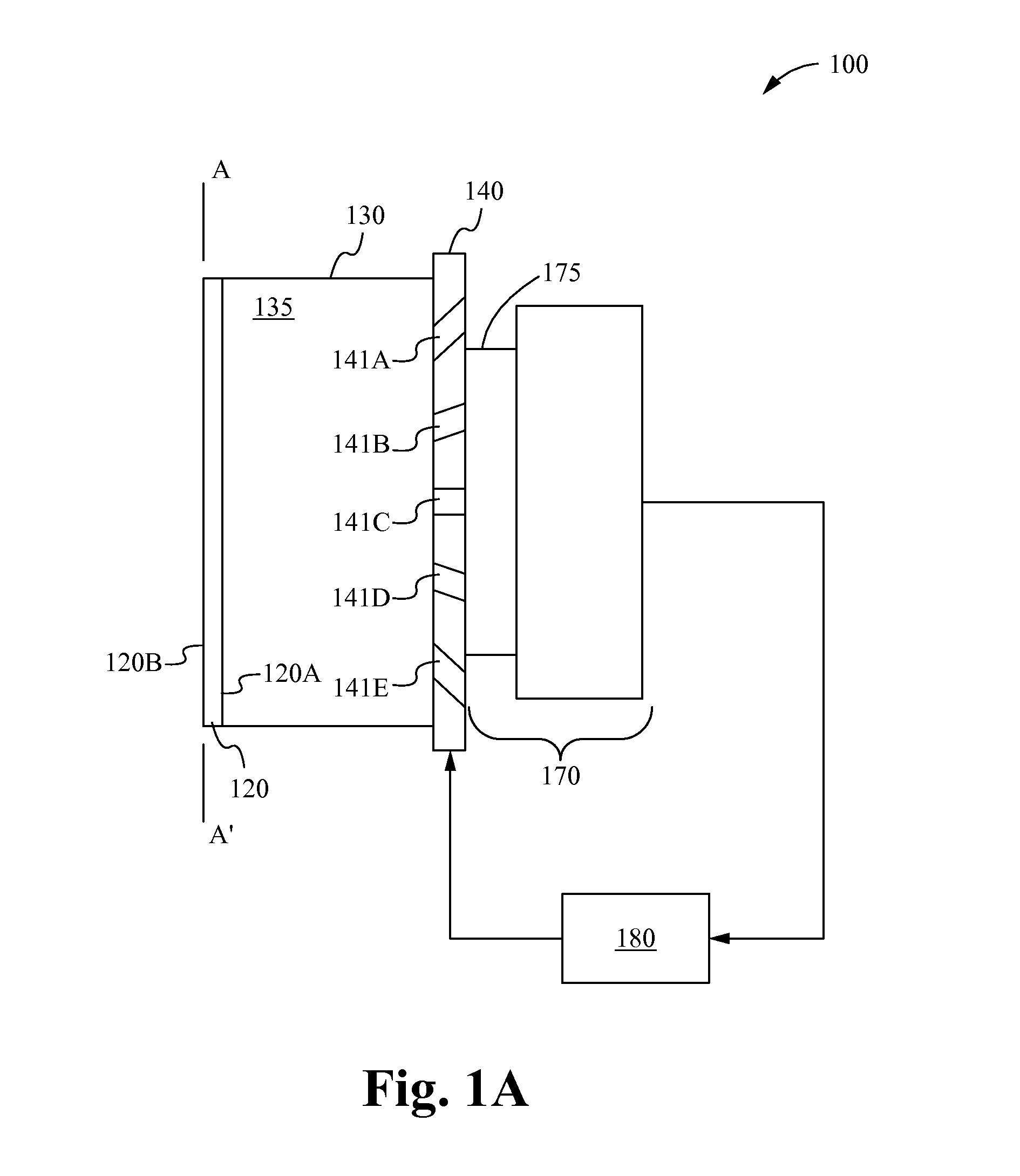
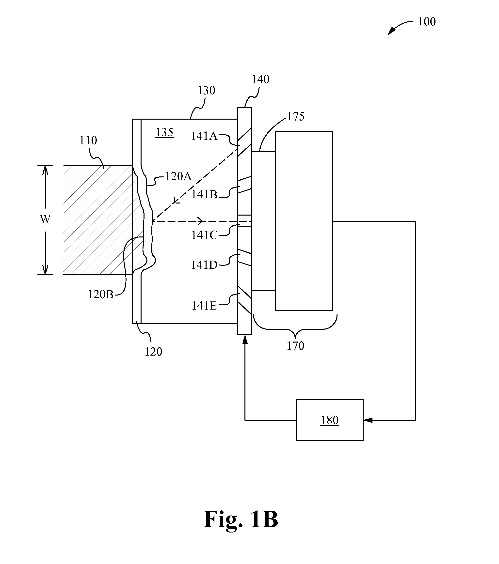
[0027]A haptic sensor in accordance with embodiments of the invention is a low-cost device that enables the real-time visualization of the haptic sense of elastic modulus boundaries, which is essentially the tissue deformation caused by a specific force. The sensor captures images that describe the three-dimensional (3-D) position and movement of underlying tissue during the application of a known force, essentially what a physician feels through manual palpation.

[0028]The sensor and supporting software enable the visualization and documentation of the equivalent of 3-D tactile input from a known applied force. The sensor eliminates the subjective analysis of physical palpation examinations and gives more accurate and repeatable results, yet is less expensive to implement than MRI, ultrasound, or similar techniques.

[0029]Data processed from captured images is also a good means for documentation for patient records. This way, physicians are also able to objectively measure change ove...


PUM


Abstract

A haptic sensor for performing palpation includes a deformable membrane having a reflective surface, a light source, a camera, and a processor. When the sensor is pressed against an object on a body, the deformable membrane deforms to contour to the shape of the object, light is reflected off the reflective surface, and captured by a camera. The reflected light is processed to reconstruct a 3-D image of the object. The rendered image can show abnormalities such as cysts, tumors, or other abnormalities, as well as arterial pressure pulses. In different embodiments, the sensor illuminates the deformed membrane from multiple directions, using shape-from-shading or grayscale mapping, or using video streams to provide more accurate images. The sensor is able to be included as part of a mobile device, such as a mobile phone, thereby making it compact and portable.

Description

CROSS REFERENCE TO RELATED APPLICATIONS[0001]This application claims priority under 35 U.S.C. §119(e) of the co-pending U.S. provisional patent application Ser. No. 61 / 577,622, filed Dec. 19, 2011, and titled “System for and Method of Quantifying On-Body Palpitation for Improved Medical Diagnosis,” which is hereby incorporated by reference.FIELD OF THE INVENTION[0002]This invention relates to object imaging. More particularly, this invention relates to reconstructing three-dimensional images from palpitations for medical purposes.BACKGROUND OF THE INVENTION[0003]Palpation is a traditional diagnostic procedure in which physicians use their fingers to externally touch and feel body tissues. Palpation is used as part of a physical examination to determine the spatial coordinates of an anatomical landmark, assess tenderness through tissue deformation, and determine the size, shape, firmness and location of an abnormality in the body through the tactile sensing of elasticity modulus diff...

Claims


Application Information

Patent Timeline
Patent Type & Authority Applications(United States)
IPC IPC(8): A61B5/00G06T7/00A61B5/11
CPCA61B5/0077A61B5/1102G06T2207/10004G06T7/0073G06T2210/41A61B5/7275A61B2576/00G06T2207/10016G06T2207/30004G01B11/24G06T7/586G16H30/40
Inventor SARRAFZADEH, MAJIDROFOUEI, MAHSANSINCLAIR, MIKE
Owner RGT UNIV OF CALIFORNIA
Learn More