Supercharge Your Innovation With Domain-Expert AI Agents!

Generating a 2D Projection Image of a Vascular System

a vascular system and projection image technology, applied in the field of generating a 2d projection image of a vascular system, can solve the problems of poor temporal resolution, restrictions on the use of three-dimensional techniques during a treatment, and the inability to eliminate the large vessels from the projection image, so as to achieve the segmentation of the large vessels easier and more accurate

Active Publication Date: 2015-07-23
SIEMENS HEALTHCARE GMBH
View PDF3 Cites 14 Cited by
  • Summary
  • Abstract
  • Description
  • Claims
  • Application Information

AI Technical Summary

Benefits of technology

The patent describes a way to make it easier to see small vessels in a 2D image of a brain. This is done by eliminating larger vessels from the image and creating a visualization of the smaller vessels in the brain, which is called a virtual parenchymogram.

Problems solved by technology

Currently, however, it is not possible to eliminate the large vessels from the projection images obtained by digital subtraction angiography.
The use of three-dimensional techniques during a treatment, however, is subject to restrictions and the temporal resolution is poorer than in the case of the 2D techniques.

Method used


Image

  • Generating a 2D Projection Image of a Vascular System
  • Generating a 2D Projection Image of a Vascular System
  • Generating a 2D Projection Image of a Vascular System

Examples


Embodiment Construction

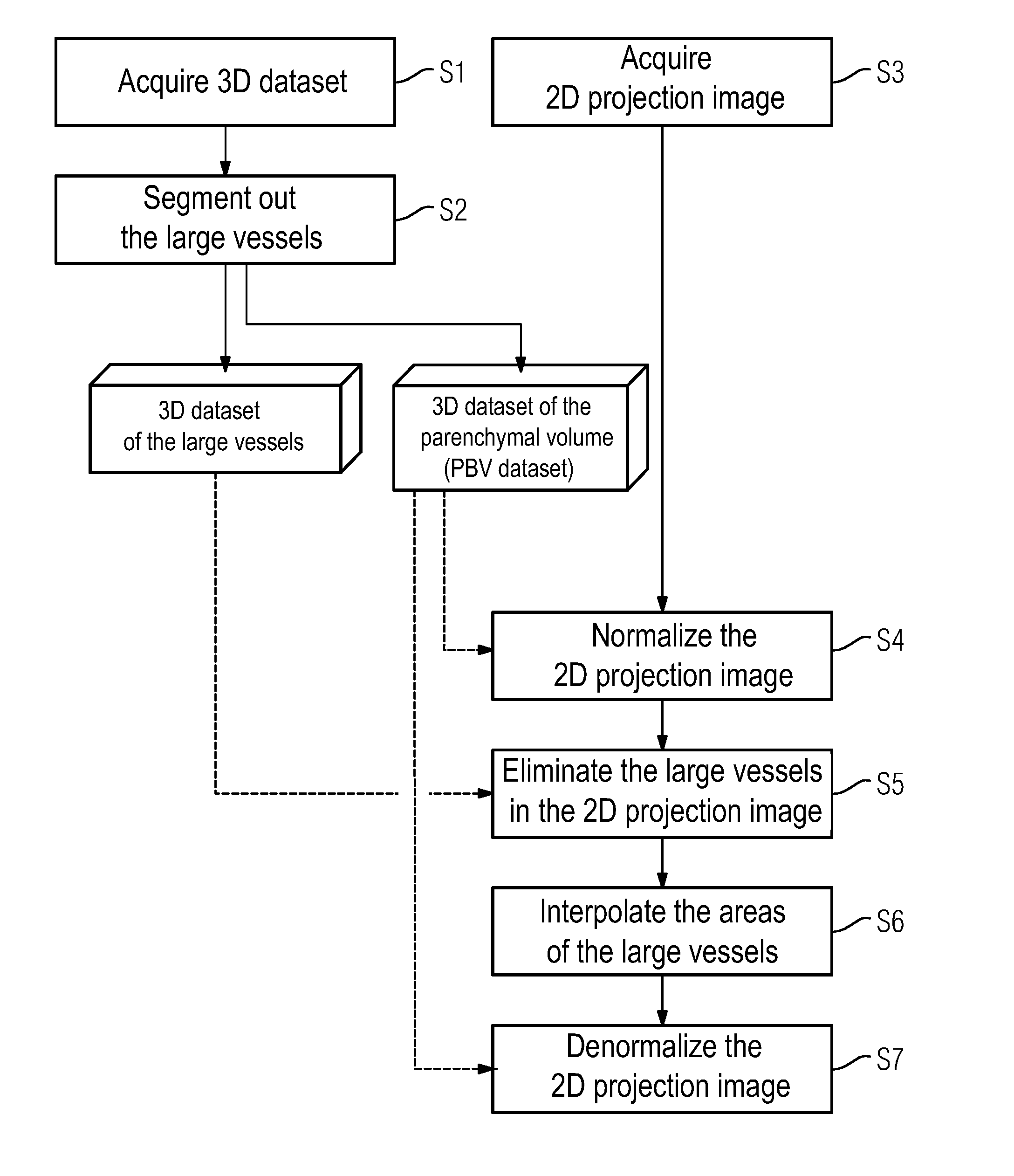
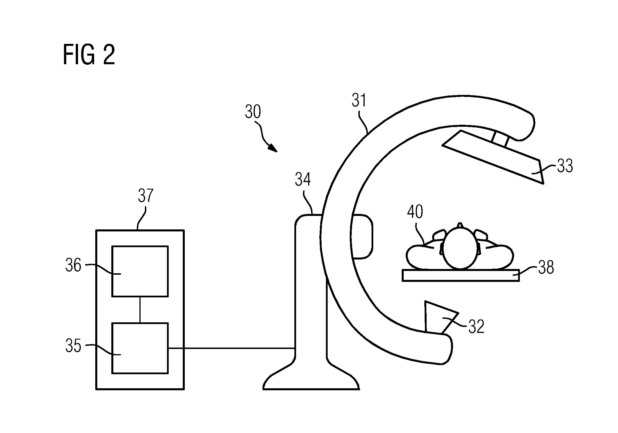
[0035]The individual method acts according to an exemplary embodiment are described below with reference to FIG. 1. In a first act S1, a 3D dataset of the vascular system is acquired by an angiography system, for example a C-arm device. The dataset may be acquired on the basis of a difference calculation between a fill image in which the vascular system is filled with contrast agent statically, e.g., uniformly or in a “steady state”, and a reference image that is free of contrast agent. The result of the 3D acquisition is a three-dimensional, static visualization of the vascular system. The acquisition contains not only the large afferent and efferent vessels (e.g., arteries, veins) but also the vessels of the parenchyma (in particular, the so-called microcirculation). All of the vessels may be segmented if necessary.

[0036]In a second act S2, those vessels whose size exceeds a predetermined limit value are segmented. This may be carried out on the basis of known segmentation methods...


PUM


Abstract

The embodiments relate to generating a 2D projection image of a vascular system of a body region of interest, including: (1) acquiring a 3D dataset of the body region of interest, (2) acquiring at least one 2D projection image of the body region of interest (S3), (3) generating a modified 3D dataset by eliminating vessels whose size exceeds a predetermined limit value, (4) normalizing the 2D projection image using projection data of the modified 3D dataset, (5) eliminating vessel projections in the normalized 2D projection image whose size exceeds a predetermined limit value, (6) interpolating the areas of the 2D projection image in which the vessel projections have been eliminated, and (7) denormalizing the normalized and interpolated 2D projection image using projection data of the modified 3D dataset.

Description

CROSS-REFERENCE TO RELATED APPLICATIONS[0001]This application claims the benefit of DE 10 2014 201 134.5, filed on Jan. 22, 2014, which is hereby incorporated by reference in its entirety.TECHNICAL FIELD[0002]The embodiments relate to a method and a device for generating at least one 2D projection image of a vascular system of a body region of interest, in particular of the parenchyma, in particular of the human or animal body.BACKGROUND[0003]Different technologies are known for visualizing a vascular system of the human or animal body. In a form of digital subtraction angiography (DSA), after administration of a contrast agent two-dimensional projection images are acquired repeatedly at a predetermined, fixed projection angle by an X-ray apparatus, a reference image (e.g., mask image) is subtracted from each image. During the procedure, the mask image is generated at a point in time at which no contrast agent is present. In this way, the arrival of the contrast agent in the vessels...

Claims


Application Information

Patent Timeline
IPC IPC(8): A61B6/00A61B5/026A61B5/00A61B6/03
CPCA61B6/5223A61B6/032A61B6/5205A61B6/504A61B6/463A61B5/7425A61B6/4441A61B5/7445A61B6/462A61B5/0263A61B5/489A61B2576/02G16H30/40
Inventor KYRIAKOU, YIANNIS
Owner SIEMENS HEALTHCARE GMBH
Features
  • R&D
  • Intellectual Property
  • Life Sciences
  • Materials
  • Tech Scout
  • Unparalleled Data Quality
  • Higher Quality Content
  • 60% Fewer Hallucinations
Learn More