Automatic background region selection for lesion delineation in medical images

a technology of background region and medical image, applied in image enhancement, instruments, applications, etc., can solve the problems of time-consuming and laborious background area creation, affecting treatment planning substantially, and prone to being defined

Active Publication Date: 2016-01-14
SIEMENS MEDICAL SOLUTIONS USA INC
View PDF13 Cites 7 Cited by
  • Summary
  • Abstract
  • Description
  • Claims
  • Application Information

AI Technical Summary

Problems solved by technology

In a typical patient medical image, a lesion area is visible in the image, but the background area is prone to being defined very subjectively and therefore inconsistently.
A lesion area would typically be visible, but the boundary with the background is typically not clear due to the resolution of the image, and image noise.
Furthermore, due to the complex shape of many background regions and the requ...

Method used


Image

  • Automatic background region selection for lesion delineation in medical images
  • Automatic background region selection for lesion delineation in medical images
  • Automatic background region selection for lesion delineation in medical images

Examples


Embodiment Construction

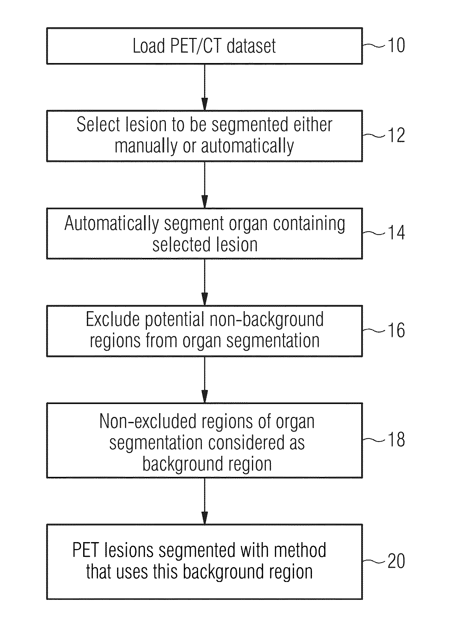
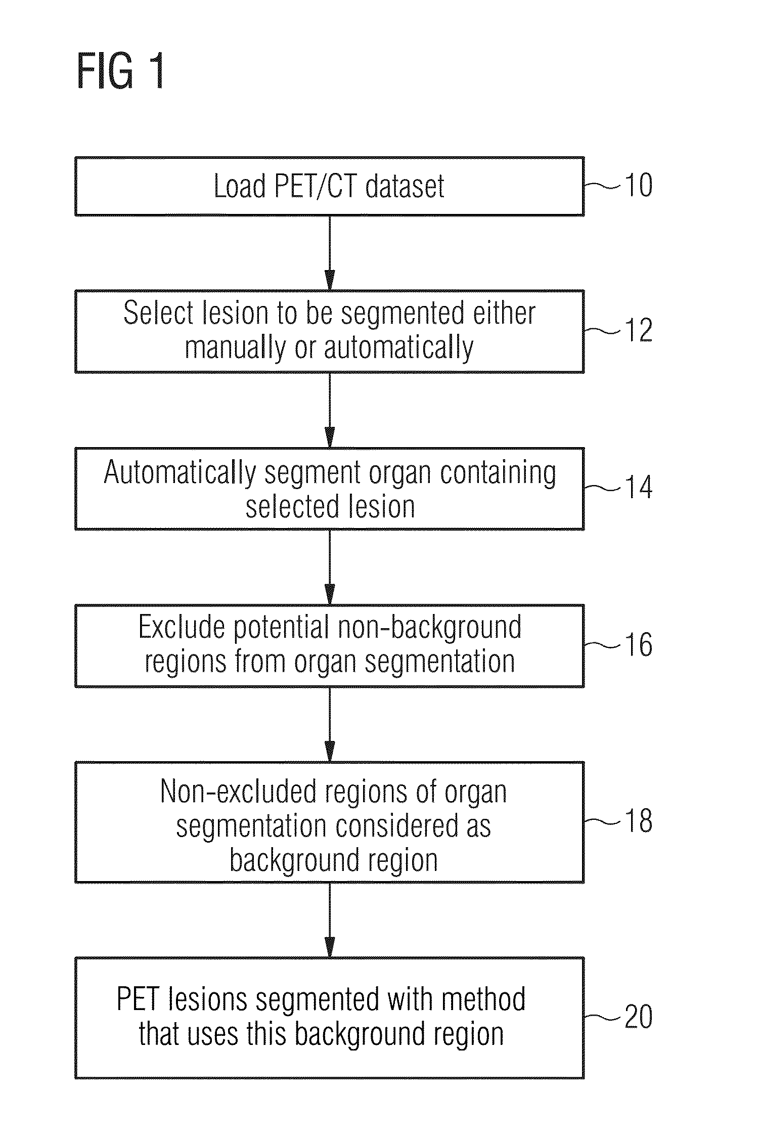
[0018]Certain embodiments of the present invention use an automated organ delineation tool which delineates an image region representing a patient organ. In an example, a co-registered PET (functional) and CT (anatomical) dataset may be used. In other examples, a single functional image data set may be used.

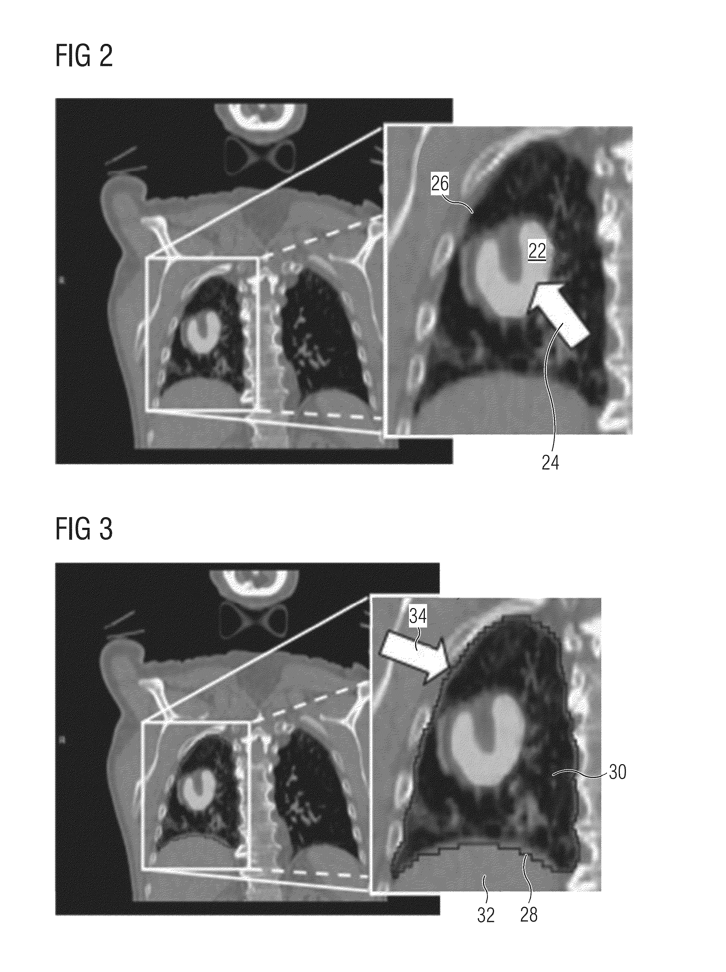
[0019]For a given target lesion, the background region is created from a delineation of an organ containing the lesion, by excluding all regions that are assumed not to be part of the background region. These regions include all representations of lesions and similar representations in terms of intensity values and shape in the organ of interest. For example, this may be defined as hotspots within a set of pre-determined SUV (standardized uptake value) thresholds in the PET image data.

[0020]In an exemplary embodiment, lung vessels or other lesions are taken to have an increased level of uptake relative to the background, as with the lesion of interest, and so can be removed from ...


PUM


Abstract

In a method and apparatus for automatic background region selection for lesion segmentation in medical images, a patient medical image dataset is loaded into a computer and an image region is delineated to obtain a segmentation containing a lesion. A background region is created from the segmentation representing the patient organ.

Description

BACKGROUND OF THE INVENTION[0001]1. Field of the Invention[0002]The present invention concerns a method and apparatus for automatically identifying a background region in order to facilitate delineation in a medical image of lesion.[0003]2. Description of the Prior Art[0004]When treating cancer using radiation or monitoring a tumor's response to the treatment, definition of gross tumor volume (GTV) is critical. GTV can be distinguished using anatomical imaging techniques such as CT or MRI. Functional imaging techniques such as PET or SPECT, on the other hand, can provide additional insight on the structure of the tumor and its metabolic activity which is used extensively as well in the treatment process. Metabolic activity may be expressed in terms of metabolic tumor volume or MTV.[0005]Accurate shape and volume determination of lesions will help clinicians to reduce radiation damage to healthy tissues surrounding the lesion and deliver maximum radiation dose to the tumor. Accurate ...

Claims


Application Information

Patent Timeline
IPC IPC(8): G06T7/00A61B5/00G06F17/30
CPCG06T7/0081A61B5/7445G06F17/30244G06T2200/04G06T2207/30096G06T2207/10104G06T2207/20144G06T2207/20148G06T2207/20104G06T2207/10081A61B5/7425A61B5/055A61B6/032A61B6/037A61B6/12A61B6/5217A61B6/5247G06T7/11G06T7/194G06T7/136G16H50/30G06T7/10G06T7/0012A61B6/52G06T7/187G06T2207/20112G06T2207/30004A61B2576/00G01T1/2992G06F16/50
Inventor FIROUZIAN, AZADEHKAFTAN, JENSKELLY, MATTHEW DAVIDMAKROPOULOS, ANTONIOS
Owner SIEMENS MEDICAL SOLUTIONS USA INC
Why Eureka
Try Eureka