Fmri-based neurologic signature of physical pain

a neurologic signature and physical pain technology, applied in the field of physical pain neurologic signature determination using fmri technology, can solve the problems of difficult to determine pain, hamper diagnosis and treatment, and burden on patients

Inactive Publication Date: 2016-02-25
THE JOHN HOPKINS UNIV SCHOOL OF MEDICINE +1
View PDF2 Cites 27 Cited by
  • Summary
  • Abstract
  • Description
  • Claims
  • Application Information

AI Technical Summary

Problems solved by technology

Physical pain is an affliction associated with enormous cognitive, social, and economic costs, but pain is not easy to ascertain.
It is primarily assessed through self-report, an imperfect measure of subjective experience, which hampers diagnosis and treatment.
The capacity to effectively report pain is limited in many vulnerable populations, such as the very old or very young, those with cognitive impairments, and those who are minimally conscious.
Moreover, self-report provides a limited basis for understanding the neurophysiological processes underlying different types of pain, and thus a limited basis for targeting treatments to the underlying neuropathology.
While application of fMRI in the context of pain is plausible, so far no reliable fMRI application to detect pain has been developed that has been demonstrated to be both sensitive and specific to pain (or any subtype of pain) within an individual person, in a manner validated across different MRI scanners.

Method used


Image

  • Fmri-based neurologic signature of physical pain
  • Fmri-based neurologic signature of physical pain
  • Fmri-based neurologic signature of physical pain

Examples


example 1

[0058]This example illustrates the methods of data acquisition and analysis used in the studies presented in Examples 2-5.

Participants

[0059]All participants provided written informed consent. Studies were individually approved by the Columbia University Institutional Review Board. For all four studies, preliminary eligibility was assessed with a general health questionnaire, a pain safety screening form, and an fMRI safety screening form. Participants reported no history of psychiatric, neurological, or pain disorders. Ethnicity was assessed using self-report screening instruments prior to study procedures.

Thermal Stimulation and Pain Rating

[0060]In all four studies, thermal stimulation was delivered to the volar surface of the left (non-dominant) inner forearm applied using a TSA-II Neurosensory Analyzer (Medoc Ltd., Chapel Hill, N.C.) with a 16 mm Peltier thermode end-plate. Each stimulus lasted 8-12 seconds, depending on the Study, and always included a period of time during whic...

example 2

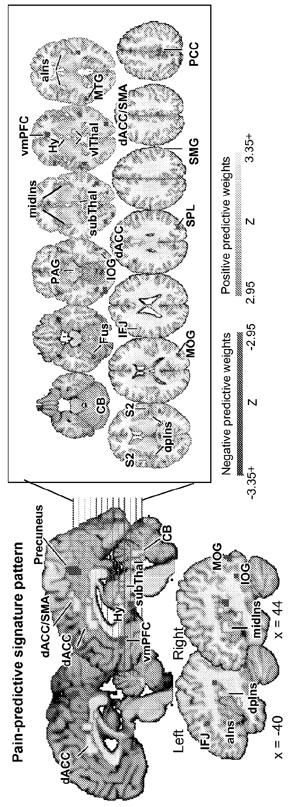
[0071]This example illustrates Study 1, which shows the development of the neurologic signature.

Participants:

[0072]Study 1 included 20 participants (aged 28.8±7.5 [S.D.] years, 8 females). The sample consisted of 79% Caucasian, 5% Hispanic, and 16% African American participants. Data were collected between 2005-2006.

Materials and Procedures:

[0073]fMRI Task Design

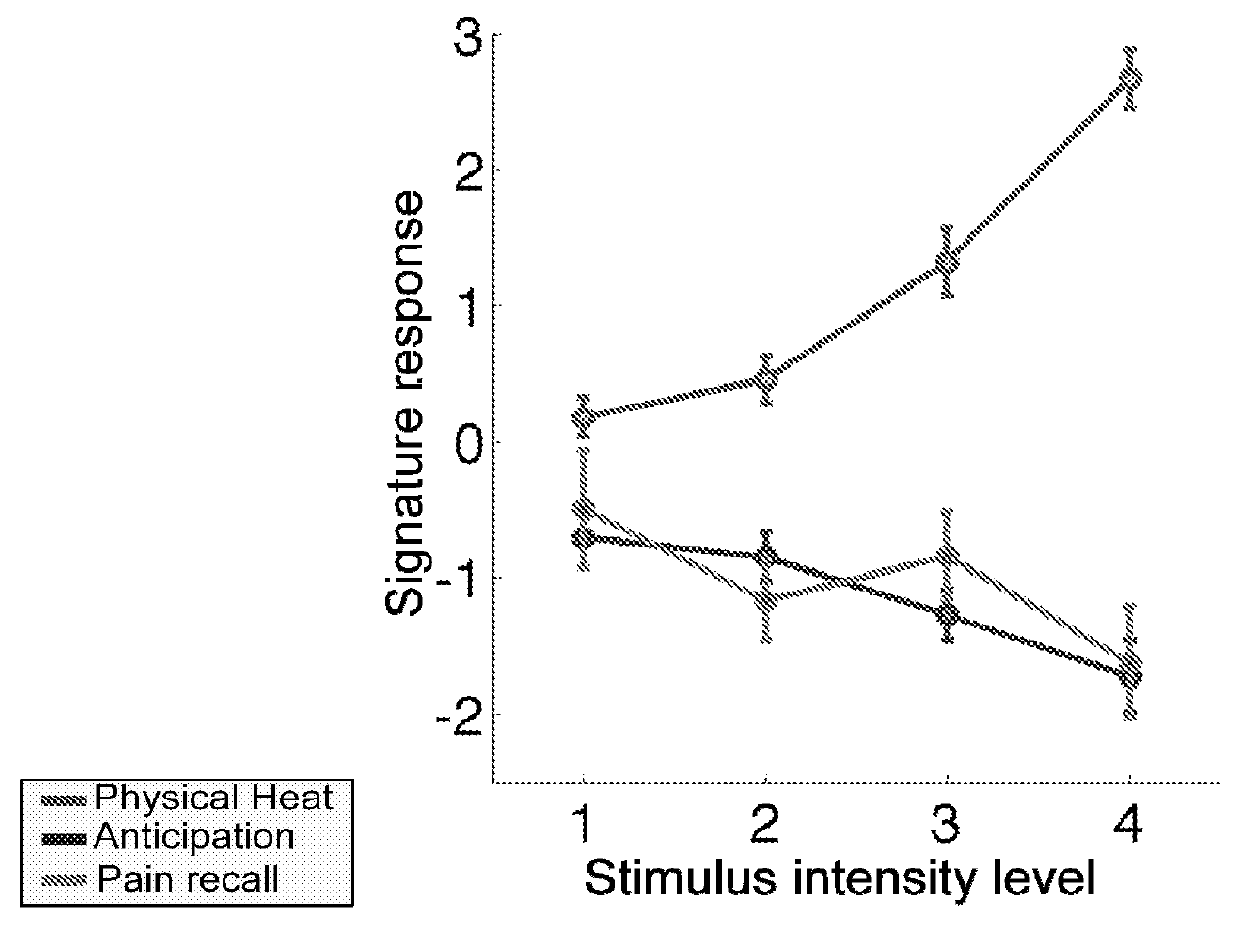
[0074]fMRI images were acquired during 8 functional runs (8 trials / run, 64 trials). The thermode was placed on a different skin site for each run, with two total runs per skin site, and 12 trials at each of 4 target pain intensities—non-painful warmth (Level 1), low pain (Level 3), medium pain (Level 5), and high pain (Level 7)—were delivered across the runs. Temperatures were selected for each individual based on a thermal pain calibration procedure (see above, “Thermal stimulation and pain ratings”). At the start of each trial, a square appeared in the center of the screen for 50 ms, followed by the presentation of a cue. ...

example 3

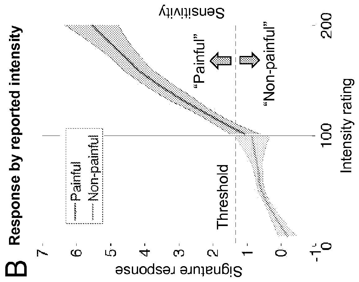
[0102]This example illustrates Study 2, which demonstrates that the neurologic signature predicts pain at the level of an individual.

Participants:

[0103]Study 2 included 33 healthy, right-handed participants (Mage=27.9±9.0 years, 22 females). The sample consisted of 39% Caucasian, 33% Asian, 12% Hispanic, and 15% African American participants.

Materials and Procedures:

Thermal Stimulation and Pain Ratings

[0104]Thermal stimulation was delivered to locations on the left volar forearm that alternated between runs. Each stimulus lasted 12.5 seconds, with 3-second ramp-up and 2-second ramp-down periods and 7.5 seconds at target temperature. Trials at six discrete temperatures were administered (level 1: 44.3° C., level 2: 45.3° C., level 3: 46.3° C., level 4: 47.3° C., level 5: 48.3° C., level 6: 49.3° C.). After each stimulus, participants rated explicitly whether it was painful or not. If they rated it as non-painful, they were then prompted to rate warmth intensity on a 100-point VAS anc...


PUM

PropertyMeasurementUnit
temperatureaaaaaaaaaa

Abstract

Described herein is a novel fMRI-based neurologic signature that predicts pain. Further described are methods for detecting pain, for diagnosing pain-related neuropathic conditions and for predicting or evaluating efficacy of an analgesic based on the neurologic signature.

Description

CROSS REFERENCE TO RELATED APPLICATION[0001]This application claims the benefit of priority under 35 U.S.C. §119(e) to U.S. Provisional Patent Application Ser. No. 61 / 810,178, filed Apr. 9, 2013, which is incorporated herein by reference.GOVERNMENT SUPPORT[0002]This invention was made with government support under grant numbers DA027794 and MH076136 awarded by the National Institutes of Health. The U.S. government has certain rights in the invention.FIELD OF THE INVENTION[0003]The present invention generally relates to the use of fMRI technology to determine a neurological signature of physical pain.BACKGROUND OF INVENTION[0004]Although biomarkers for medical conditions have proliferated over the past 50 years, objective assessments related to mental health have lagged behind. Physical pain is an affliction associated with enormous cognitive, social, and economic costs, but pain is not easy to ascertain. It is primarily assessed through self-report, an imperfect measure of subjectiv...

Claims


Application Information

Patent Timeline
Patent Type & Authority Applications(United States)
IPC IPC(8): G01R33/48A61B5/00A61B5/055G01N33/49
CPCG01R33/4806A61B5/055A61B5/4824A61B2576/026A61B5/4848G01N33/4925A61B5/4839
Inventor WAGER, TORLINDQUIST, MARTIN
Owner THE JOHN HOPKINS UNIV SCHOOL OF MEDICINE
Why Eureka
Try Eureka