
Reducing Vibration of a Cantilevered Patient Support Surface

a patient support surface and cantilever technology, applied in the field of system for reducing vibration of, can solve the problem of small vibration of the patient support surface, and achieve the effect of reducing vibration of the cantilever patient support surfa

Active Publication Date: 2016-06-30
GENERAL ELECTRIC CO
View PDF1 Cites 0 Cited by
  • Summary
  • Abstract
  • Description
  • Claims
  • Application Information

AI Technical Summary

Benefits of technology

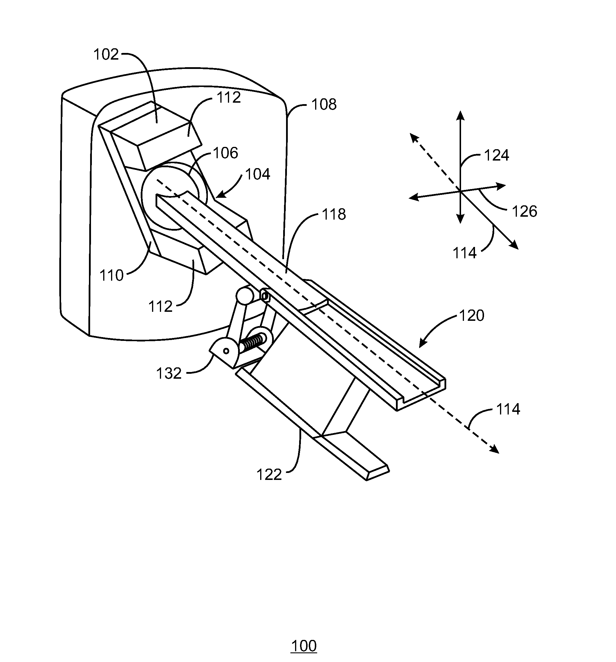
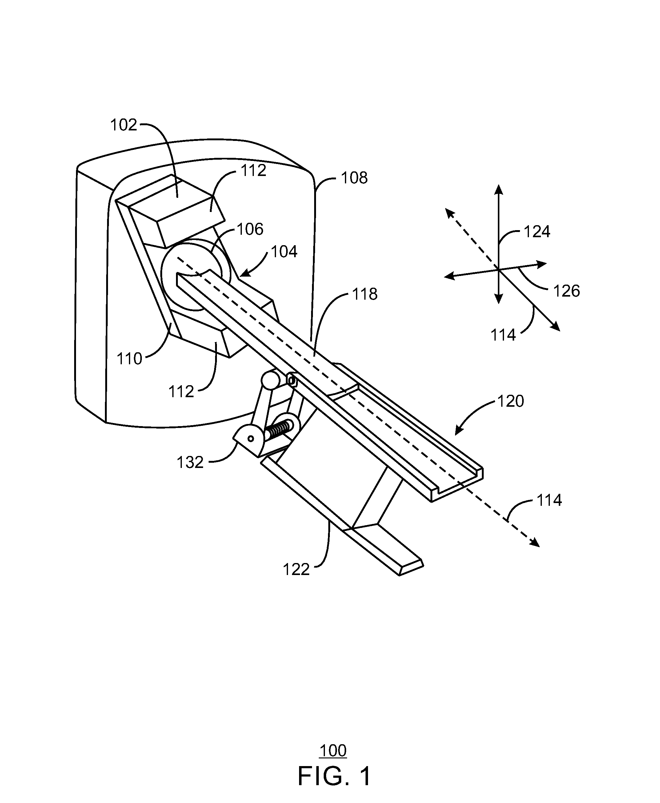
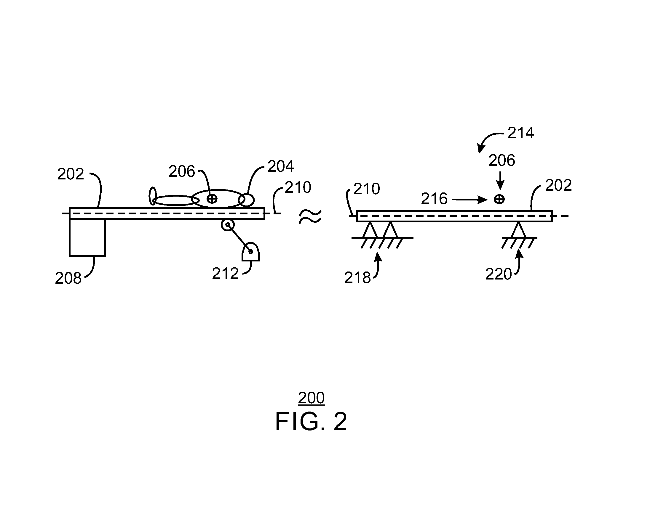
This patent is about a system and method to make a patient support surface less vibrant. The system includes a spring and roller that work together to keep the surface stable and prevent bouncing. This technology ensures that the patient's position stays stable during medical imaging without causing any movement.

Problems solved by technology

The movement of the patient to obtain PET scans and CT scans can create small vibrations that can create artifacts within an image obtained from a PET or CT scan.

Method used


Image

  • Reducing Vibration of a Cantilevered Patient Support Surface
  • Reducing Vibration of a Cantilevered Patient Support Surface
  • Reducing Vibration of a Cantilevered Patient Support Surface

Examples


Embodiment Construction

[0013]In the following detailed description, reference is made to the accompanying drawings that form a part hereof, and in which is shown by way of illustration specific embodiments that may be practiced. These embodiments are described in sufficient detail to enable those skilled in the art to practice the embodiments, and it is to be understood that other embodiments may be utilized and that logical, mechanical, electrical and other changes may be made without departing from the scope of the embodiments. The following detailed description is, therefore, not to be taken as limiting the scope of the invention.

[0014]As discussed above, a patient is supported by a patient support surface and then moved within the gantry on the patient support surface to obtain PET scans and / or CT scans of the patient's anatomy. As used herein, the patient support surface includes, but is not limited to, a cradle movably attached to a table support. In the embodiments discussed herein, a device is use...


PUM


Abstract

A system and method for reducing vibration of a cantilevered patient support surface are provided herein. A system includes a spring and roller coupled with the spring. The spring creates an upwards spring bias on the patient support surface. The roller is coupled with the spring via a roller lever arm. A frictional resistance at the roller arm and an upward directed spring bias enables the roller to maintain constant contact with the patient support surface.

Description

BACKGROUND OF THE INVENTION[0001]Various technologies exist wherein portions of a patient are imaged to study tissues or organs within the body. For example, nuclear medicine includes techniques where a radiopharmaceutical is introduced to a patient that causes the emission of photons from the body of the patient. The radiopharmaceutical may concentrate in particular tissues of the body, indicating tissue metabolic activity at the site of concentration and emitting a higher amount of photons from the site of concentration. Images may be reconstructed from photons observed during Positron Emission Tomography (PET) scan. PET scans may be combined with computerized tomography (CT) scans to develop an image of tissues and organs of a patient.[0002]A patient is supported by a patient support surface and then moved within a gantry on the patient support surface to obtain PET scans and CT scans of the patient's anatomy. The movement of the patient to obtain PET scans and CT scans can creat...

Claims


Application Information

Patent Timeline
Patent Type & Authority Applications(United States)
IPC IPC(8): F16F15/02F16M11/22A61B6/04
CPCF16F15/022F16M11/22A61B6/0428A61B6/032A61B6/037A61B6/0407F16F15/06F16F1/06F16F15/067
Inventor GRAMS, ROBERT SCOTTOLIKARA, PHILIPNATHAN, ADAM CLARK
Owner GENERAL ELECTRIC CO
Learn More