Medical image processing apparatus and medical image processing method

a medical image and processing apparatus technology, applied in the field of medical image processing methods and apparatuses, can solve the problems of great concern in the manipulation of the mri apparatus, and achieve the effect of convenient user manipulation

Inactive Publication Date: 2016-08-04
SAMSUNG ELECTRONICS CO LTD
View PDF4 Cites 3 Cited by
  • Summary
  • Abstract
  • Description
  • Claims
  • Application Information

AI Technical Summary

Benefits of technology

[0008]The present disclosure provides methods and apparatuses for processing a medical image that facilitate convenient user manipulation.
[0009]The present disclosure also provides methods and apparatuses for processing a medical image which provide for a contour correction of an object where a segmentation result may be corrected via a smaller number of user inputs.
[0013]The processor may decrease the displayed size of the endo-contour image and increase the displayed size of the epi-contour image in response to the first user input, or increase the displayed size of the endo-contour image and decrease the displayed size of the epi-contour image in response to the first user input.
[0023]The processor may decrease the displayed size of a first contour image from among the plurality of contour images and / or increase the displayed size of a second contour image from among the plurality of contour images in response to the first user input, or increase the displayed size of the first contour image and decrease the size of the second contour image in response to the first user input.

Problems solved by technology

Since the user is engaged in repeatedly manipulating the MRI apparatus over the entire life of the MRI apparatus, convenient manipulation of the MRI apparatus is an issue of great concern.

Method used


Image

  • Medical image processing apparatus and medical image processing method
  • Medical image processing apparatus and medical image processing method
  • Medical image processing apparatus and medical image processing method

Examples


Embodiment Construction

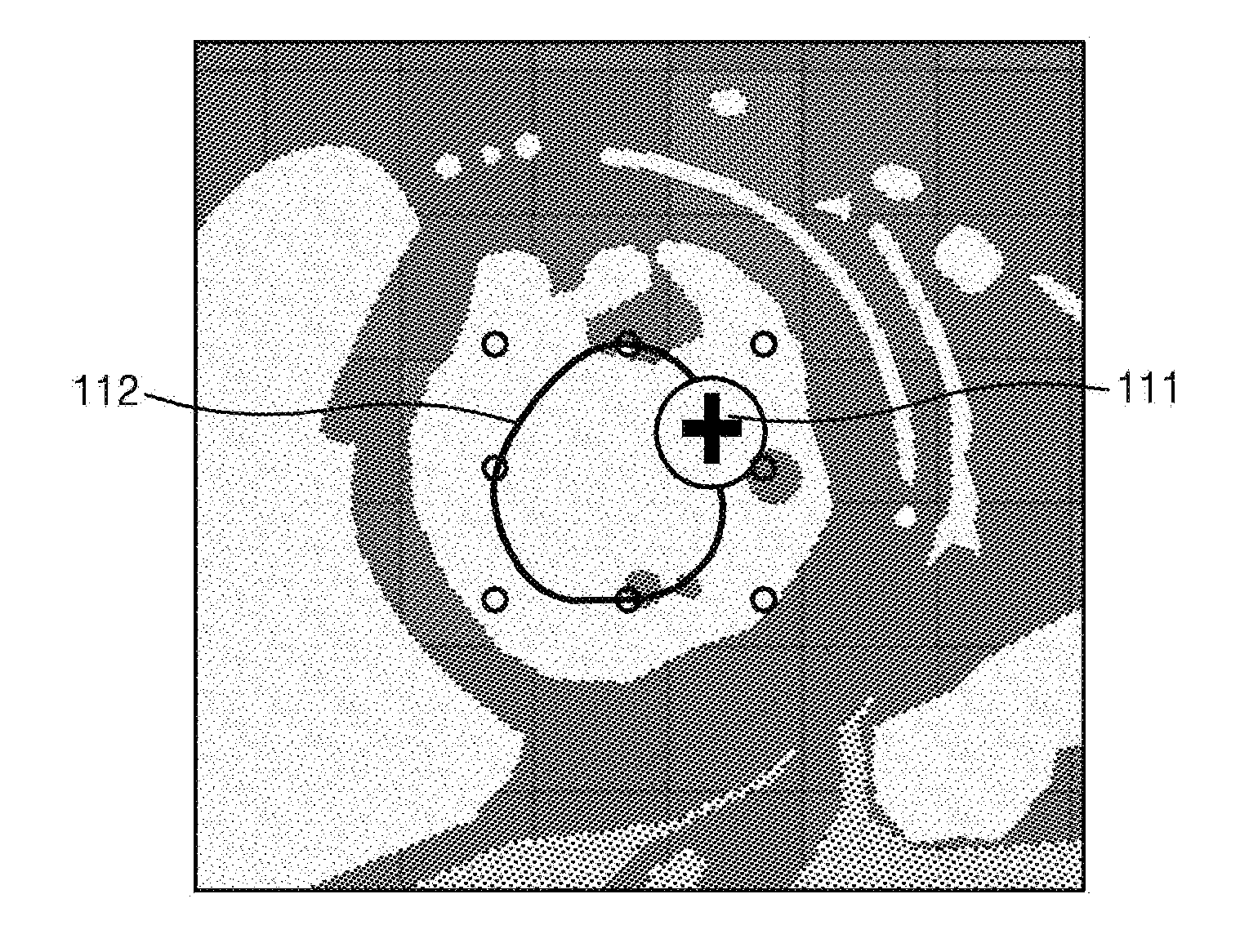
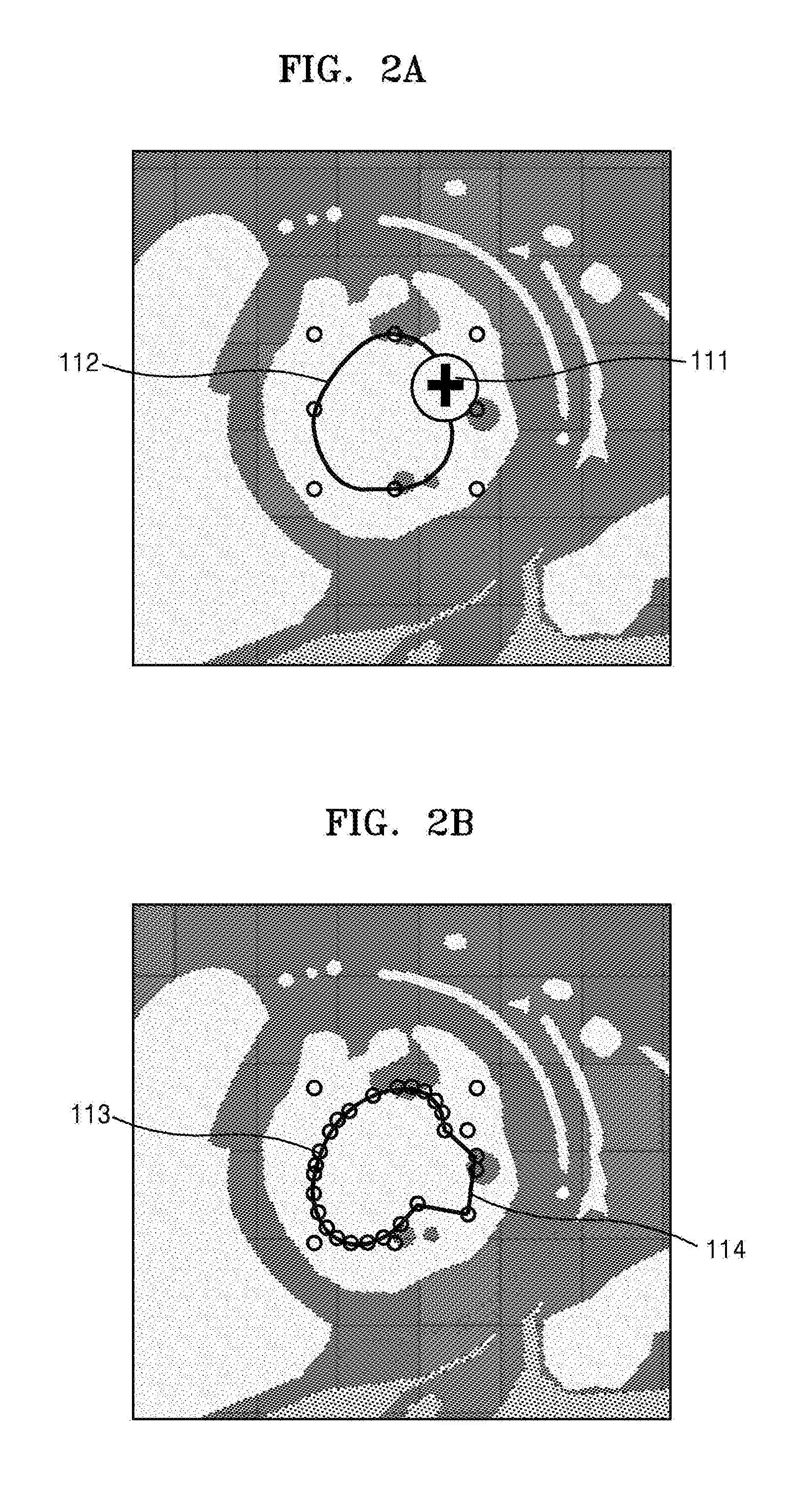
[0067]Advantages and features of the one or more embodiments of the present disclosure and methods of accomplishing the same may be understood more readily by reference to the following detailed description of the embodiments and the accompanying drawings. In this regard, the present embodiments may have different forms and should not be construed as being limited to the descriptions set forth herein. Rather, these embodiments are provided so that this disclosure will be thorough and complete in terms of understanding and supporting the claimed subject matter and will fully convey the concept of the present embodiments to one of ordinary skill in the art, as the present disclosure will only be defined by the appended claims.

[0068]Terms used herein will now be briefly described and then one or more embodiments of the present disclosure will be described in detail.

[0069]All terms including descriptive or technical terms which are used herein should be construed as having meanings that...


PUM


Abstract

A medical image processing apparatus provides a contour correction of an object where a segmentation result is correctable via a smaller number of user inputs. The apparatus includes: a display screen configured to display an endo-contour image and an epi-contour image corresponding to a myocardial image; an input interface configured to receive a first user input for the endo-contour image and epi-contour image. A processor is configured to change the displayed size of the endo-contour image and epi-contour image in response to the first user input, wherein the display outputs a display of both of the changed endo-contour image and epi-contour image together.

Description

CLAIM OF PRIORITY[0001]This application claims the benefit of Korean Patent Application No. 10-2015-0014585, filed on Jan. 29, 2015, in the Korean Intellectual Property Office, the disclosure of which is incorporated herein in its entirety by reference.BACKGROUND[0002]1. Field of the Disclosure[0003]The present disclosure relates to methods and apparatuses for processing a medical image. More particularly, the present disclosure relates to methods and apparatuses for processing a medical image which are capable of providing a user with a convenient editing environment.[0004]2. Description of the Related Art[0005]Medical imaging apparatuses are used to acquire images showing an internal structure of an object, typically a living being or a portion of the living being. Medical imaging apparatuses are non-invasive examination apparatuses that capture and process images of details of structures, tissue, fluid flow, etc., inside a body and provide the images to a user. A user, e.g., a me...

Claims


Application Information

Patent Timeline
Patent Type & Authority Applications(United States)
IPC IPC(8): G06F3/0484G06T7/00
CPCG06F3/04847G06T2200/24G06T7/0012G06F3/04845G06F2203/04806G06T2207/20096G06T2207/30048G06T7/12A61B5/0044A61B5/055A61B5/742A61B5/7475
Inventor JO, HYUN-HEEPARK, SEON-MI
Owner SAMSUNG ELECTRONICS CO LTD
Why Eureka
Try Eureka