Methods and systems for automatic segmentation

a technology of automatic segmentation and image, applied in the field of non-invasive diagnostic imaging, can solve the problems of fbp's suboptimal noise and image quality performance, many segmentation algorithms are often distracted, and many segmentation algorithms fail to identify correct organ boundaries, etc., to achieve accurate segmentation, improve the reliability and robustness of the segmentation algorithm, and save the textural details of the image.

Inactive Publication Date: 2016-10-06
GENERAL ELECTRIC CO
View PDF4 Cites 10 Cited by
  • Summary
  • Abstract
  • Description
  • Claims
  • Application Information

AI Technical Summary

Benefits of technology

[0004]In one embodiment, a method comprises generating a first image from acquired projection data based on an iterative reconstruction algorithm, generating a second image from the acquired projection data based on a modified iterative reconstruction algorithm, segmenting the second image to obtain segments, segmenting the first image based on the segments of the second image, and outputting the segmented

Problems solved by technology

Unfortunately, many segmentation algorithms are often distracted by the presence of noise and fail to identify correct organ boundaries.
However, one disadvantage of FBP is its suboptimal noise and image quality performance res

Method used


Image

  • Methods and systems for automatic segmentation
  • Methods and systems for automatic segmentation
  • Methods and systems for automatic segmentation

Examples


Embodiment Construction

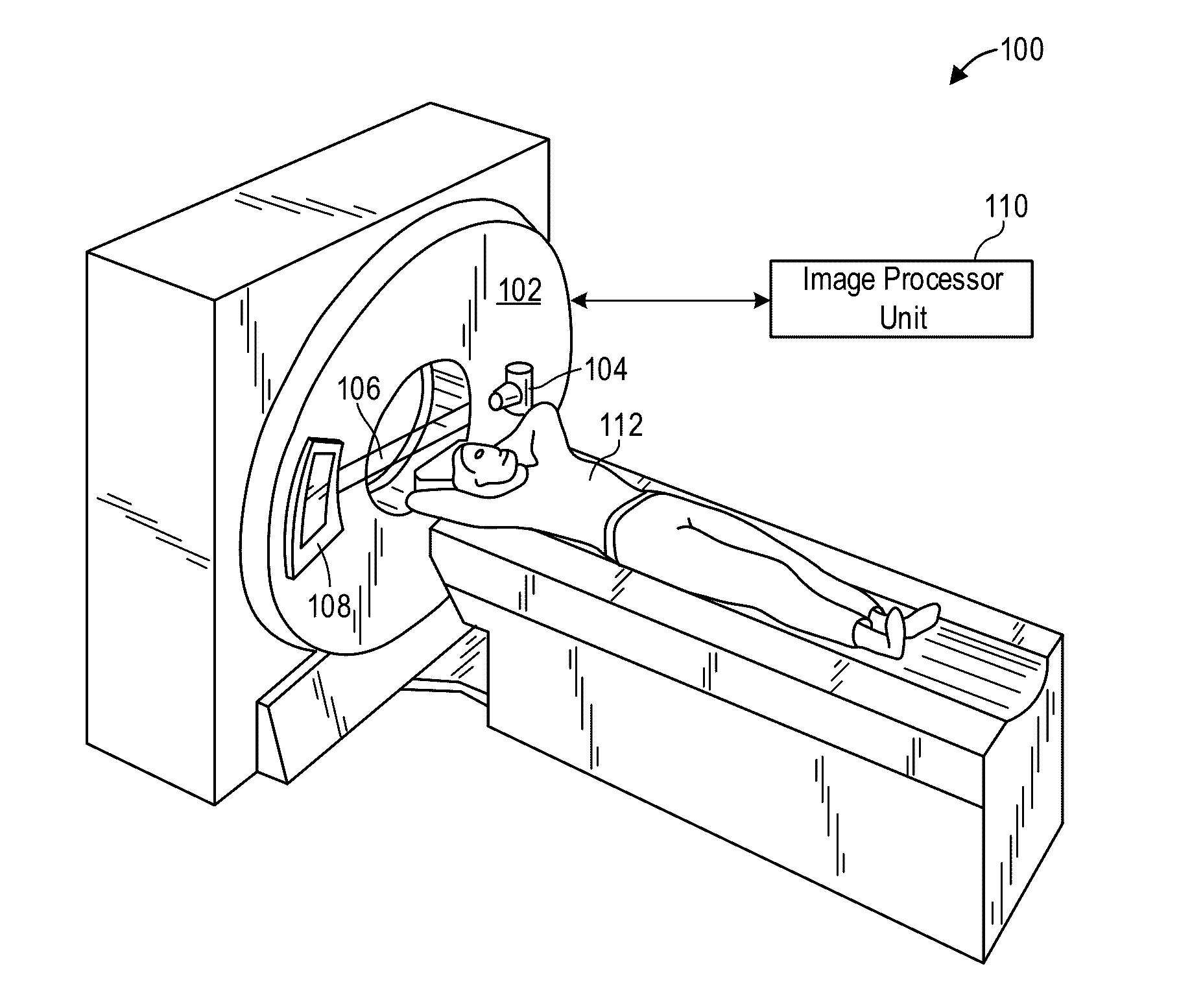
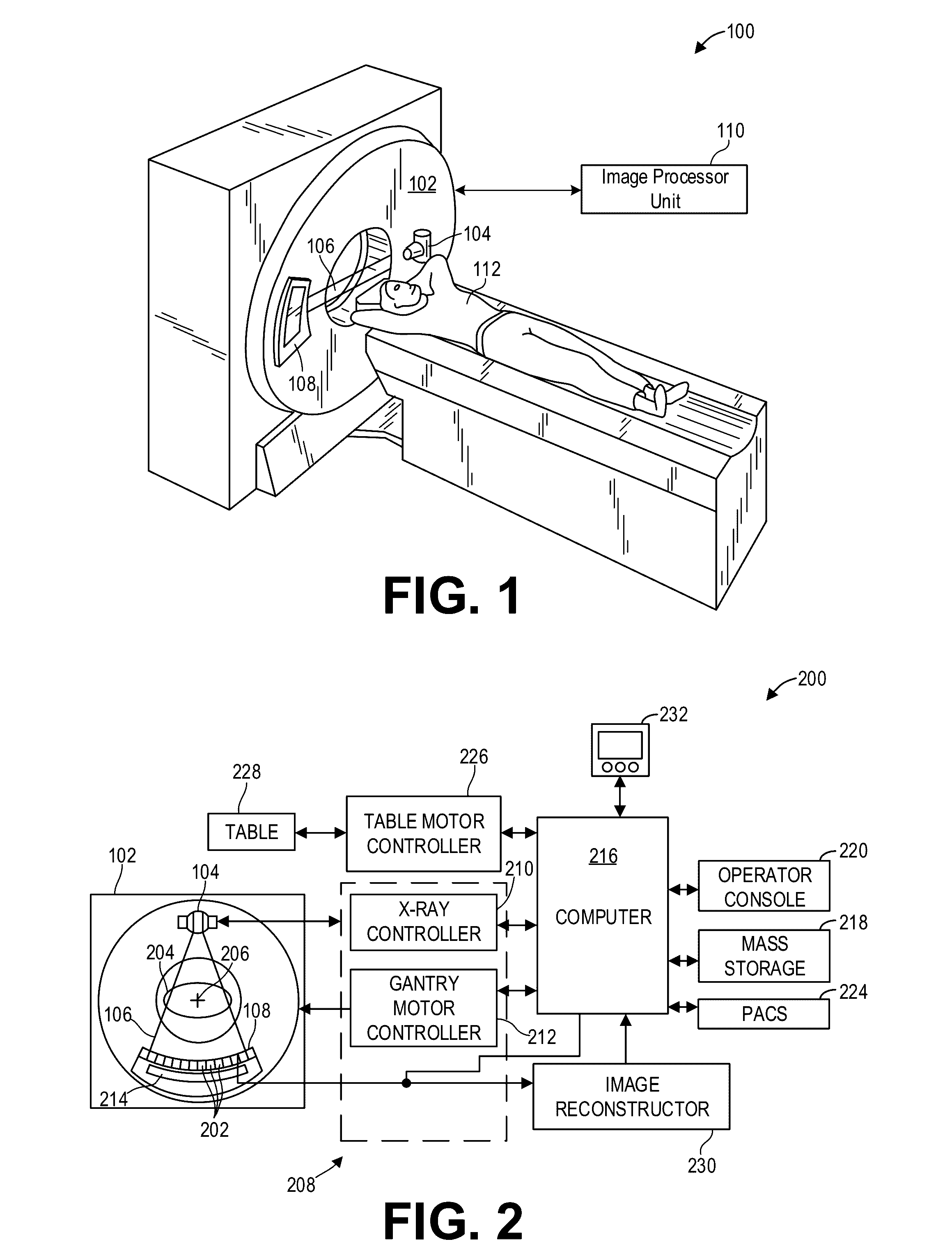
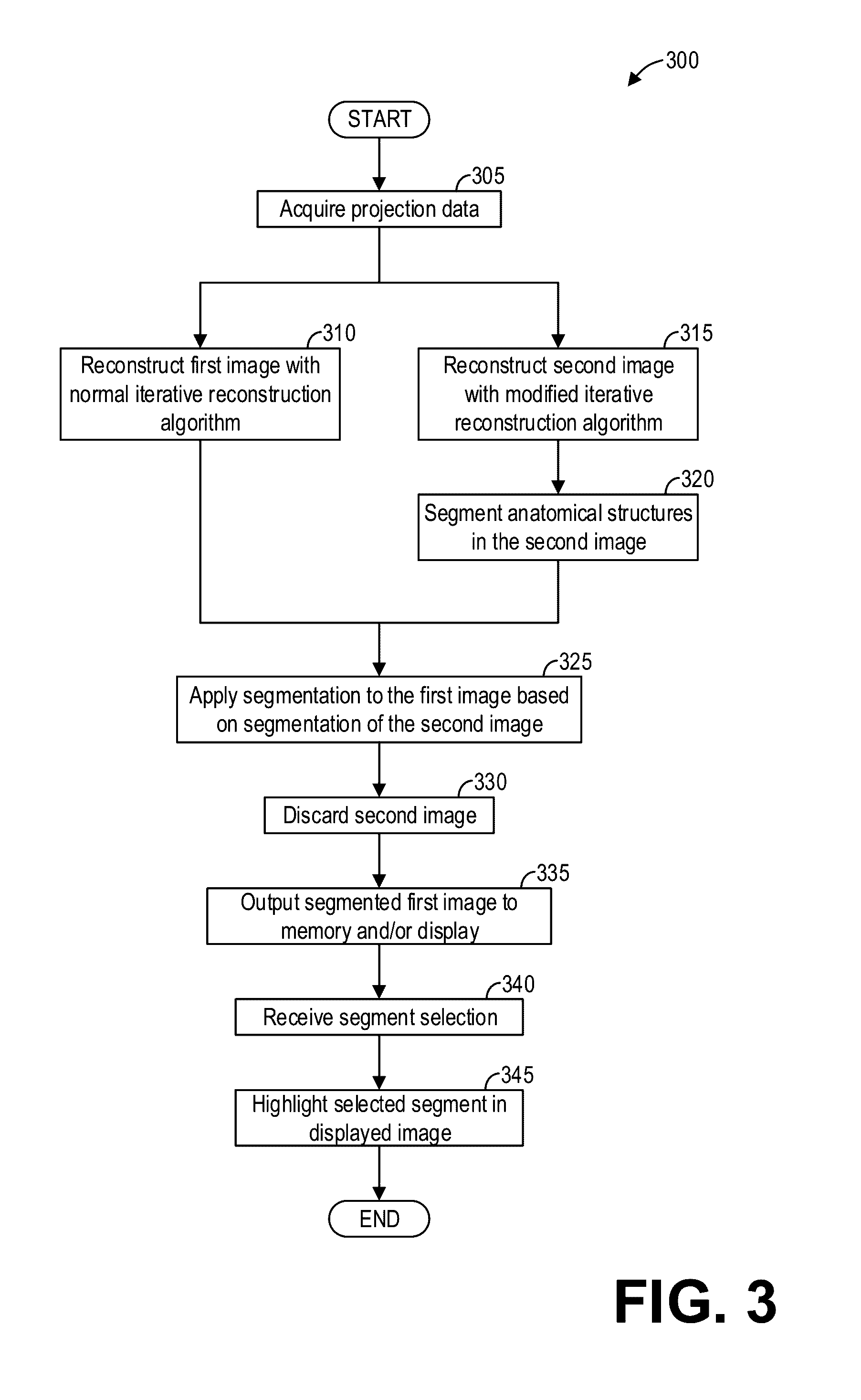
[0013]The following description relates to various embodiments of medical imaging systems. In particular, methods and systems are provided for reconstructing an image with ideal properties for automatic segmentation. An example of a computed tomography (CT) imaging system that may be used to acquire images processed in accordance with the present techniques is provided in FIGS. 1 and 2. A method for automatic segmentation, such as the method shown in FIG. 3, may include reconstructing a first image suitable for diagnostics and a second image suitable for segmentation, and the first image may be segmented based on the segmentation of the second image. FIGS. 4 and 5 show example image reconstructions suitable for diagnostics and segmentation, respectively. When the segmentation of an image takes significant priority over the textural details of an image, a method for automatic segmentation, such as the method shown in FIG. 6, may include reconstructing an image suitable for segmentati...


PUM


Abstract

Methods and systems are provided for reconstructing and automatically segmenting an image. In one embodiment, a method comprises generating a first image from acquired projection data based on an iterative reconstruction algorithm, generating a second image from the acquired projection data based on a modified iterative reconstruction algorithm, segmenting the second image to obtain segments, segmenting the first image based on the segments of the second image, and outputting the segmented first image to a display. In this way, an image which may otherwise prove challenging for an automatic segmentation process may be accurately segmented without sacrificing textural details of the image.

Description

FIELD[0001]Embodiments of the subject matter disclosed herein relate to non-invasive diagnostic imaging, and more particularly, to automatic segmentation of diagnostic images.BACKGROUND[0002]Non-invasive imaging technologies allow images of the internal structures of a patient or object to be obtained without performing an invasive procedure on the patient or object. In particular, technologies such as computed tomography (CT) use various physical principals, such as the differential transmission of x-rays through the target volume, to acquire image data and to construct tomographic images (e.g., three-dimensional representations of the interior of the human body or of other imaged structures).[0003]One of the key tasks in oncology is to automatically perform segmentation of reconstructed images to identify organs and other anatomical structures. Unfortunately, many segmentation algorithms are often distracted by the presence of noise and fail to identify correct organ boundaries. F...

Claims


Application Information

Patent Timeline
IPC IPC(8): G06T7/00G06T11/00
CPCG06T7/0079G06T2207/10004G06T2207/10081G06T11/003G06T2207/30004G06T7/11G06T7/174G06T2211/424G06T11/008
Inventor HSIEH, JIANGDOAN, WILLIAM DAVIDANDERSON, PAUL ROGER
Owner GENERAL ELECTRIC CO
Why Eureka
Try Eureka