Method for 2D/3D Registration, Computational Apparatus, and Computer Program

a technology of computational apparatus and computer program, applied in image enhancement, tomography, instruments, etc., can solve problems such as inability to accurately superimpose, inability to accurately predict movement, and interruption of the person carrying out the procedur

Active Publication Date: 2016-11-17
SIEMENS HEALTHCARE GMBH
View PDF8 Cites 13 Cited by
  • Summary
  • Abstract
  • Description
  • Claims
  • Application Information

AI Technical Summary

Benefits of technology

[0014]The embodiments are based on the object of specifying a registration method, a computational apparatus and a computer program, which permit a robust and accurate registration, even when proceeding from less exact 2D / 3D correspondences and / or, in particular, over a relatively long series of recorded x-ray images.
[0020]Overall, the method consequently reduces the computational outlay by virtue of mapping the reference structures as a sparse representation, consequently focusing on the initial points and the contour points associated therewith. The comparison method is carried out locally. Once a 2D / 3D correspondence has been found locally, it may not be lost in further iterations. In the proposed approach, a gradient comparison image is generated directly from the three-dimensional image data record, with the search region for the 2D / 3D tracking possibly being based on the image content, namely—as will still be explained in more detail below—on the direction of the gradient in the three-dimensional image data record. In this way, the method provides valid registrations both at the start of a medical procedure and also during the further course thereof, and also during the recording breaks for the x-ray images; after the three-dimensional image data record is used as an initial point as a matter of principle, there also is no further accumulation of tracking errors, since an accurate registration between the three-dimensional image data record and the two-dimensional x-ray image is established immediately. In a certain way, the proposed method therefore provides a natural (re-)initialization for the differential movement compensation, particularly if the initial transformation is based on the registration transformation established at the previous time step. In this way, the movement compensation framework becomes more reliable since no errors accumulate and a refinement of the registration ultimately occurs automatically when it becomes necessary. Persons carrying out a medical procedure may concentrate more on the course of the procedure, without being interrupted by incorrect superpositions and the like.
[0022]It was found that a suitable initial transformation may already be established with sufficient accuracy from a known position of the patient in relation to the x-ray apparatus recording the x-ray image. When positioning patients within an x-ray apparatus having, e.g., a C-arm, in particular for a medical procedure, the location of the patient, and consequently the position of the target region, may be already known very accurately, with, furthermore, the recording geometry and the geometric properties of the x-ray apparatus likewise being available such that, in the case of a known recording geometry, the initial transformation may be estimated with sufficient accuracy in order to lead to sufficiently reliable success in the 2D / 3D registration, in particular in the case of a suitable parameterization of the registration algorithm proposed here, in particular in respect of the extent of the neighborhoods and / or the search region.
[0024]As already mentioned above, the present method is suitable, in particular, for fluoroscopic x-ray monitoring of, in particular, minimally invasive medical procedures. In this case, it is particularly expedient if a time series of x-ray images is recorded, in particular for monitoring a medical procedure, wherein each x-ray image is registered and the registration transformation established in each case for the previously immediately recorded x-ray image is used as an initial transformation. Since there may be only a short time interval between the recording of temporally successive x-ray images, particularly strong movements are not expected in the target region, and so the final registration transformation related to the previously recorded x-ray image is particularly well suited as an initial transformation for the next registration procedure. A quality of the initial transformation known in this manner may also be used, as described above, to adapt parameters, in particular in view of the described 3D / 2D tracking, such that one example provides for a smaller neighborhood and / or a smaller number of test points to be selected in the case of a registration transformation as an initial transformation determined for an x-ray image recorded immediately therebefore than in the case of any other initial transformation. By way of example, if the x-ray image has a resolution of 512*512 pixel, a neighborhood with dimensions of 20*20 pixel may be formulated in the case of a large assumed deviation of the initial transformation, in particular when first determining a registration transformation from an approximate transformation, e.g., at the outset, wherein the neighborhood used in comparison may be reduced to 5*5 pixel when using registration transformations of preceding x-ray images. Similarly, the number of test points or the search region may be selected to be smaller.
[0032]Consequently, a particular refinement provides for, in addition to a base test point corresponding to the contour point, test points to be selected along the direction of the gradient, projected onto the image plane, at the initial point associated with the contour point. In other words, the three-dimensional gradients are taken into account when the search direction for finding correspondences is set. Here, this is an intuitive search direction as this projection of the three-dimensional gradient ultimately specifies precisely the direction in which the movements are observable. Accordingly, there may also be a targeted search in this case, the outlay of which is significantly reduced and contributes to the real-time capability of the described procedure. Ultimately, the search direction is selected on the basis of the image content instead of in a gradient-based manner in an optimization method, as is conventional.
[0041]What emerges from the considerations presented above is that the two-dimensional and three-dimensional movements are linked to one another by the target plane π, which contains the target point of the movement w+dw (e.g., the new three-dimensional position of the initial point W after the movement), the projection center C (e.g., the focus of the radiation source, which may be selected as the origin of the coordinate system) and the moved two-dimensional contour point p+dp, e.g., the comparison point p′. What emerges from these geometric considerations is that it is possible to determine a normal vector of the target plane π, which may be used as a computational means.

Problems solved by technology

However, the 2D / 3D registration may become invalid during the medical procedure due to movements of the patient, e.g., of the target region; consequently, incorrect superpositions may occur.
A disadvantage here is that the person carrying out the procedure is interrupted when carrying out the interventional procedure by the movement correction.
However, the error occurring in the movement estimation is disadvantageously identifiable in the fusion or superposition images after the tracking errors have accumulated and 2D / 3D correspondences are lost by the movement.
Despite all these approaches, the 2D / 3D registration still continues to pose a challenge, in particular when it is intended to be carried out in real time.
However, the optimization is made more difficult in this case by the loss of the depth information occurring as a result of the projection.
Another category of known methods uses the back projection of the x-ray image and compares the latter with the three-dimensional image data record, with these approaches, however, requiring a plurality of two-dimensional x-ray images recorded under different projection directions, e.g., in different recording geometries, which is often not the case in medical procedures.
However, the problems of accumulating tracking errors and also the possible loss of 2D / 3D correspondences also occur in this case.

Method used


Image

  • Method for 2D/3D Registration, Computational Apparatus, and Computer Program
  • Method for 2D/3D Registration, Computational Apparatus, and Computer Program
  • Method for 2D/3D Registration, Computational Apparatus, and Computer Program

Examples


Embodiment Construction

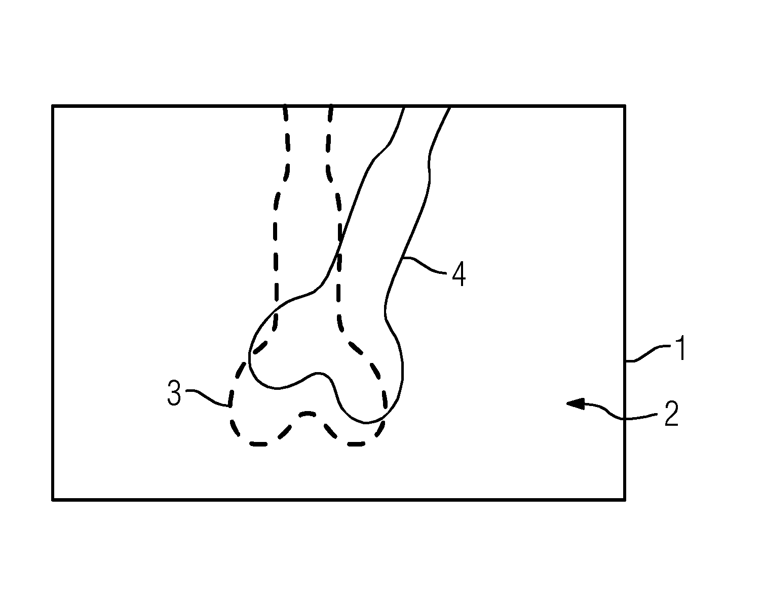
[0062]FIG. 1 depicts, in an exemplary manner and in principle, a superposition image 1 made of a three-dimensional image data record and an x-ray image, wherein the x-ray image 2 forms the basis on which information 3 from the three-dimensional image data record is superposed. An anatomical structure 4, in this case a bone, is identifiable in the x-ray image 2 in a shadow-like manner and with a low resolution. The information 3 relating to the same anatomical structure 4 was superposed from the three-dimensional image data record, wherein the superposition was brought about using an approximate initial transformation, and consequently an approximate 2D / 3D registration. The anatomical structure in accordance with the information 3 is slightly twisted and translated in relation to the visible anatomical structure 4 of the x-ray image 2. The exemplary embodiment of the method described below is now directed to establishing a registration transformation that leads to an accurate superpo...


PUM


Abstract

A method for registering a three-dimensional image data record of a target region of a patient with a two-dimensional x-ray image is provided. The method includes selecting at least one rigid reference structure with an associated contour; establishing a two-dimensional gradient x-ray image and a three-dimensional gradient data record of the image data record; finding a neighborhood in the gradient x-ray image from a plurality of neighborhoods extending about test points for a plurality of contour points; establishing local two-dimensional displacement information by comparison of the contour points with the associated comparison points; establishing movement parameters of a three-dimensional movement model describing a movement of the target region between recording of the image data record and the x-ray image from the local two-dimensional displacement information; and establishing a registration transformation describing the registration by correcting the test transformation based on the movement parameters.

Description

[0001]This application claims the benefit of DE 10 2015 208 929.0, filed on May 13, 2015, which is hereby incorporated by reference in its entirety.TECHNICAL FIELD[0002]The embodiments relate to a method for registering a three-dimensional image data record of a target region of a patient with a two-dimensional x-ray image of the target region recorded in a recording geometry, a computational apparatus, and a computer program.BACKGROUND[0003]Both three-dimensional image data in the form of an image data record, for example a computed tomography or magnetic resonance data record, and two-dimensional fluoroscopy images, e.g., x-ray images, of the target region of interest in the patient are available in many medical problems—be it in diagnostics or when carrying out minimally invasive procedures in particular. Here, it is desirable to be able to evaluate both the three-dimensional image data record and the two-dimensional x-ray image of the target region together, in particular, in th...

Claims


Application Information

Patent Timeline
Patent Type & Authority Applications(United States)
IPC IPC(8): G06T7/00G06F19/00G16H30/20G16H30/40
CPCG06T7/0083G06T7/0012G06T2207/10028G06T2207/10116G06T2207/30008G06F19/321A61B2090/364A61B6/032A61B6/4441A61B6/487A61B6/5223A61B6/5235G06T2207/10072G06T2207/10124G06T7/33G06T7/269A61B2090/376G16H30/40G16H30/20A61B90/00G06T7/12
Inventor BORSDORF, ANJAWANG, JIAN
Owner SIEMENS HEALTHCARE GMBH
Why Eureka
Try Eureka