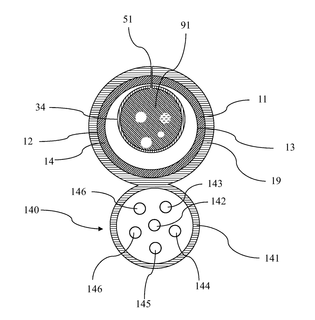
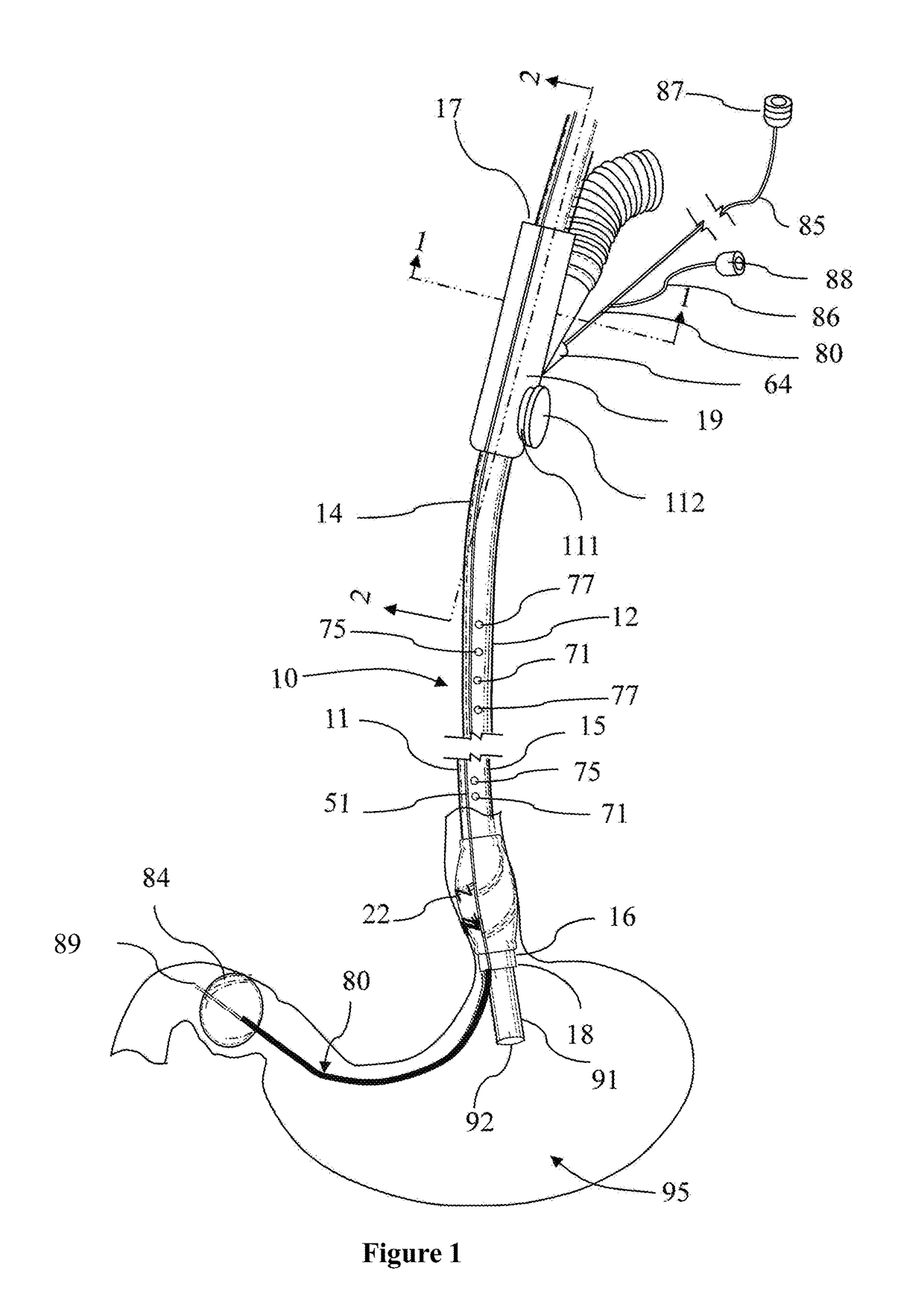
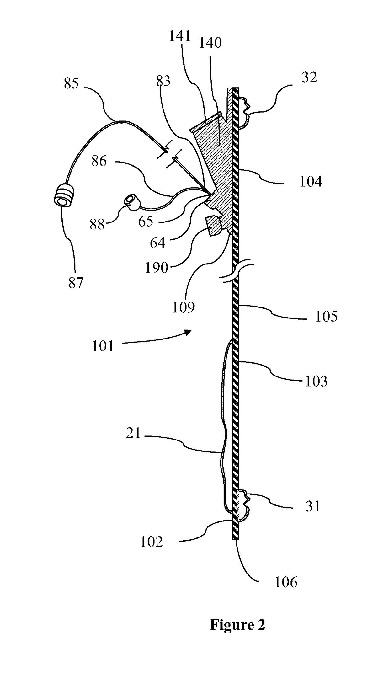
Endoscope Accessory

a technology of endoscope and accessory, which is applied in the field of endoscope accessories, can solve the problems of affecting the obtaining of ultrasonic images, affecting the patient's bowel movement, so as to improve the endoscopic examination and enhance the luminal view

Active Publication Date: 2017-03-09
IZOMED INC
View PDF0 Cites 12 Cited by
  • Summary
  • Abstract
  • Description
  • Claims
  • Application Information

AI Technical Summary

Benefits of technology

[0011]Some embodiments of the present invention improves endoscopic examination by enhancing and maintaining luminal view by deploying the proposed device. The endoscopic examination method comprises placing an endoscope shaft within a body cavity at a desired examination point, the endoscope shaft having a proximal end, mid shaft and a distal end, the distal end terminating in an endoscope tip, wherein the endoscope tip is proximal to the desired examination point, positioning a segment of a longitudinally opened overtube over the endoscope mid shaft, wherein the overtube including a longitudinal reclosable seam along an entire length of the overtube, an inner surface, an outer surface, a proximal end and a distal end, a positioning ring adjacent the distal end on the outer surface, at least one sealing band on the inner surface, and an independently positionable occlusion catheter terminating in an asymmetrical occlusion balloon; at least one handle at the proximal end and on the outer surface for grasping and manipulation of the overtube within the body cavity. The method further comprises closing the seam of the segment of the overtube positioned over the endoscope mid shaft to

Problems solved by technology

This can cause bloating and discomfort to the patient or, in rare cases, severe abdominal pain.
Air, however, creates a barrier to ultrasonic wave passage and hampers the obtaining of ultrasonic images.
In most parts of the gastrointestinal tract, however, the large diameter of the lumen and/or the angle of the transducer in relation to the intestinal wall results in an inadequate contact between the transducer balloon and the intestinal wall.
A significant shortcoming of this prior art is that it does not account for the fact that the gastrointestinal tract is not a closed region and the infused

Method used


Image

  • Endoscope Accessory
  • Endoscope Accessory
  • Endoscope Accessory

Examples


Embodiment Construction

[0093]Before explaining some aspects of embodiment of the present invention in detail, it is to be understood that the present invention is not limited in its application to the details of arrangements of the components set forth in the following description. As can be appreciated by those skilled in the arts, the present invention is capable of other embodiments and of being practiced and carried out in various ways. Also, it is to be understood that the phraseology and terminology employed herein are for the purpose of description and should not be regarded as limiting. It is also to be understood that where ranges are provided for various aspects of the invention and for examples, they are approximate ranges and are not to be limiting except where noted otherwise.

[0094]Unless defined otherwise, all technical and scientific terms used herein have the same meaning as commonly understood by one of ordinary skill in the art to which this invention belongs. Moreover, the singular form...


PUM


Abstract

This invention relates generally to a method for performing an endoscopic procedure.
According to some aspects of the invention, the method comprises placing an endoscope shaft within a body cavity at a desired examination point, the endoscope shaft having a proximal end, mid shaft and a distal end, the distal end terminating in an endoscope tip, wherein the endoscope tip is proximal to the desired examination point, positioning a segment of a longitudinally opened overtube over the endoscope mid shaft, wherein the overtube including a longitudinal reclosable seam along an entire length of the overtube, an inner surface, an outer surface, a proximal end and a distal end, a positioning ring adjacent the distal end on the outer surface, at least one sealing band on the inner surface, and an independently positionable occlusion catheter terminating in an asymmetrical occlusion balloon; at least one handle at the proximal end and on the outer surface for grasping and manipulation of the overtube within the body cavity. The method further comprises closing the seam of the segment of the overtube positioned over the endoscope mid shaft to form a longitudinally closed overtube and move the closed overtube over the endoscope shaft as guide within the body cavity and repeat the closing and moving till the overtube reaches the desired examination point, inflating the positioning ring to create a seal between the outer surface of the overtube and a body cavity proximal to the endoscope tip, wherein when inflated the positioning ring expanded asymmetrically around the overtube, inflating the at least one sealing band to create a seal between the internal surface of the overtube and endoscope shaft, passing the independently positionable occlusion catheter terminating in the asymmetrical occlusion balloon through a passageway along the overtube to enter the body cavity at the end of the overtube, manipulating the independently positionable occlusion catheter to a selected point within the body cavity distal to the endoscope tip, inflating the occlusion catheter balloon to create seal between the occlusion catheter balloon and the body cavity, and creating a sealed examination compartment between the positioning ring, the asymmetrical occlusion balloon and the sealing band surrounding the distal end of the endoscope shaft.

Description

CROSS-REFERENCE TO RELATED APPLICATIONS[0001]This application is a continuation-in-part of U.S. Ser. No. 14 / 575,509 filed on Dec. 18, 2014 and Ser. No. 14 / 121,563 filed on Sep. 18, 2014 and U.S. Ser. No. 13 / 900,524 filed on May 22, 2013 which is a continuation-in-part of U.S. Ser. No. 12 / 266,953 filed on Nov. 7, 2008, both of which are incorporated herein by reference.I. FIELD OF INVENTION[0002]Some embodiments of the present invention relates generally to the field of endoscopy and, more particularly, to an endoscope accessory for improving endoscopic examination of body organs, particularly the gastrointestinal tract.II. BACKGROUND OF INVENTION[0003]Endoscopy is a well-known procedure for examining the internal organs. The procedure is performed under the guidance of an endoscope. Currently used fiber optic endoscopes include lenses mounted in a flexible tube that relay an image from inside a body cavity for viewing by a physician for diagnosis or manipulation inside the body cavi...

Claims


Application Information

Patent Timeline
IPC IPC(8): A61B1/00
CPCA61B1/00154A61B1/00135A61B1/00082A61B1/015
Inventor FARHADI, ASHKAN
Owner IZOMED INC
Why Eureka
Try Eureka