Nanoscale Imaging of Proteins and Nucleic Acids via Expansion Microscopy

a technology of expansion microscopy and protein, applied in the field of nanoscale imaging of proteins and nucleic acids via expansion microscopy, can solve the problems of inability to image rna in intact tissues, inability to retain native proteins in gels, and inability to image genetically encoded fluorophores without antibody labeling

Active Publication Date: 2017-03-09
PRESIDENT & FELLOWS OF HARVARD COLLEGE +1
View PDF1 Cites 98 Cited by
  • Summary
  • Abstract
  • Description
  • Claims
  • Application Information

AI Technical Summary

Benefits of technology

[0005]A small molecule linker is synthesized that enables RNA to be covalently attached to the ExM gel. This method, referred to as ExFISH, enables RNA fluorescent in situ hybridization (FISH), which enables identification of transcripts in situ with single molecule precision. In RNA FISH, a set of fluorescent probes complementary to a target strand of mRNA are delivered2,3. Single molecule FISH (smFISH) can be performed with multiple fluorophores delivered to a single mRNA via oligonucleotide probes4. In intact tissues, amplific

Problems solved by technology

However, it has remained difficult to image RNA in intact tissues with the nanoscale precision required to pinpoint associations with cellular compartments or proteins important for RNA function.
Another drawback of t

Method used


Image

  • Nanoscale Imaging of Proteins and Nucleic Acids via Expansion Microscopy
  • Nanoscale Imaging of Proteins and Nucleic Acids via Expansion Microscopy
  • Nanoscale Imaging of Proteins and Nucleic Acids via Expansion Microscopy

Examples


Embodiment Construction

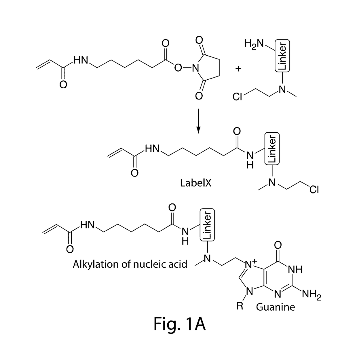
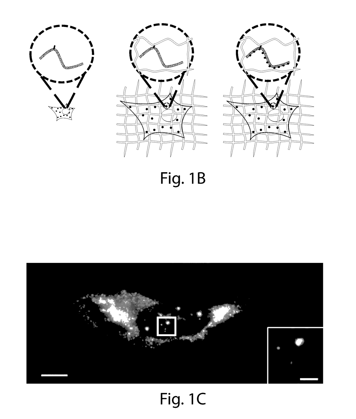
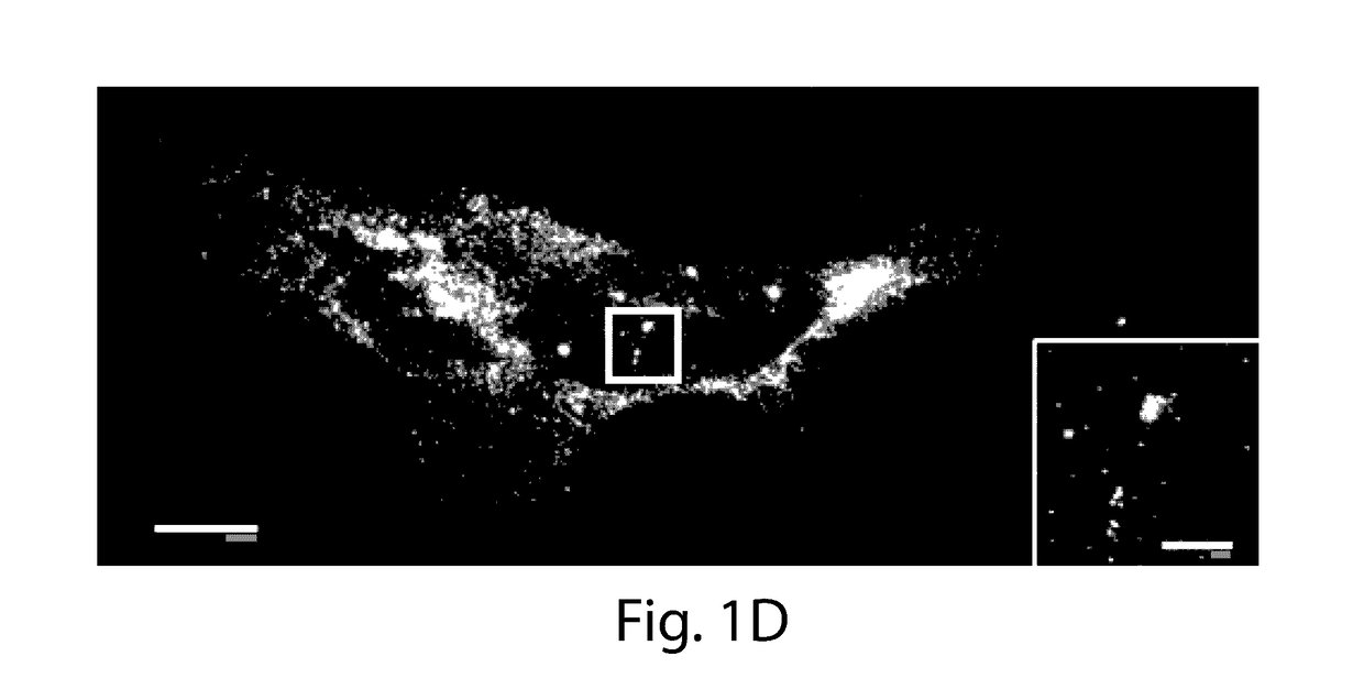
[0023]The present invention provides for the anchoring of nucleic acids into the swellable gel of Expansion Microscopy (ExM), both for in site genomic and transcriptomic assessment, as well as to enable nucleic acid barcodes to be used to identify essentially arbitrary numbers of molecules. International patent application serial number PCT / US15 / 16788, which is incorporated herein by reference, teaches that the resolution of conventional microscopy can be increased by physically expanding specimens, a process termed ‘expansion microscopy’ (ExM). In short, biological specimens are embedded in a swellable gel material, subjected to a treatment to disrupt native biological networks, and then expanded. The advantages to ExM include tissue clearing, resolution improvement, and higher tolerance to sectioning error due to the specimen expansion in the z-axis.

[0024]In ExM, fluorophores were anchored directly to the polymer gel, so that proteins could be visualized; however, RNA molecules we...


PUM

PropertyMeasurementUnit
Fractionaaaaaaaaaa
Temperatureaaaaaaaaaa

Abstract

The invention enables in situ genomic and transcriptomic assessment of nucleic acids to be conducted in biological specimens that have been physically expanded. The invention leverages the techniques for expansion microscopy (ExM) to provide new methods for in situ genomic and transcriptomic assessment of nucleic in a new process referred to herein as “expansion fluorescent in situ hybridization” (ExFISH).

Description

RELATED APPLICATION[0001]This application claims the benefit of U.S. Provisional Application Ser. No. 62 / 202,421, filed Aug. 7, 2015, the contents of which are incorporated herein by reference in its entirety.GOVERNMENT SUPPORT[0002]This invention was made with government support under 5-DPI-NS087724 awarded by NIH, Hertz Foundation, ODGE Lemelson & Viterbi, 5-DPI-N S087724 awarded by NIH and NSF. The government has certain rights in the invention.BACKGROUND OF THE INVENTION[0003]Nanoscale-resolution imaging of RNA throughout cells, tissues, and organs is key for an understanding of local RNA processing, mapping structural roles of RNA, and defining cell types and states. However, it has remained difficult to image RNA in intact tissues with the nanoscale precision required to pinpoint associations with cellular compartments or proteins important for RNA function.[0004]Expansion microscopy (ExM) enables imaging of thick preserved specimens with ˜70 nm lateral resolution. Using ExM t...

Claims


Application Information

Patent Timeline
IPC IPC(8): C12Q1/68
CPCC12Q1/6841C12Q1/6806G01N1/36G01N2001/364
Inventor WASSIE, ASMAMAWCHEN, FEIBOYDEN, EDWARD STUARTALON, SHAHAR
Owner PRESIDENT & FELLOWS OF HARVARD COLLEGE
Why Eureka
Try Eureka