Retention and stabilization of anatomy for ultrasound imaging

a technology of anatomy and ultrasound imaging, applied in mammography, medical science, diagnostics, etc., can solve the problems of patient motion, image artifacts in medical imaging systems, blurry imaging, etc., and achieve the effect of minimizing imaging artifacts

Inactive Publication Date: 2017-04-27
QT ULTRASOUND
View PDF6 Cites 1 Cited by
  • Summary
  • Abstract
  • Description
  • Claims
  • Application Information

AI Technical Summary

Benefits of technology

[0002]Systems and methods for retention and stabilization of anatomy for ultrasound imaging are described. In some cases, body parts can be stabilized against a flexible membrane for patient comfort and minimizing imaging artifacts by using pressurized fluid that holds the body part against the flexible membrane during a medical imaging procedure.

Problems solved by technology

Patient motion is the single largest cause of image artifacts in medical imaging systems.
As these medical imaging systems are developed to achieve higher and higher resolutions, even very small patient motions can blur the imaging results.
Physically restraining the patient can be very uncomfortable for the patient.
For sensitive body parts, this can be a particularly difficult issue as people will tend to postpone or simply not do important medical imaging screening tests like mammography to avoid patient discomfort.

Method used


Image

  • Retention and stabilization of anatomy for ultrasound imaging
  • Retention and stabilization of anatomy for ultrasound imaging
  • Retention and stabilization of anatomy for ultrasound imaging

Examples


Embodiment Construction

[0015]Systems and methods for the retention and stabilization of anatomy for ultrasound imaging are described. The described system and methods can be a valuable tool in the stabilization of body parts for comfort positioning during medical imaging procedures and for minimizing the occurrence of imaging artifacts.

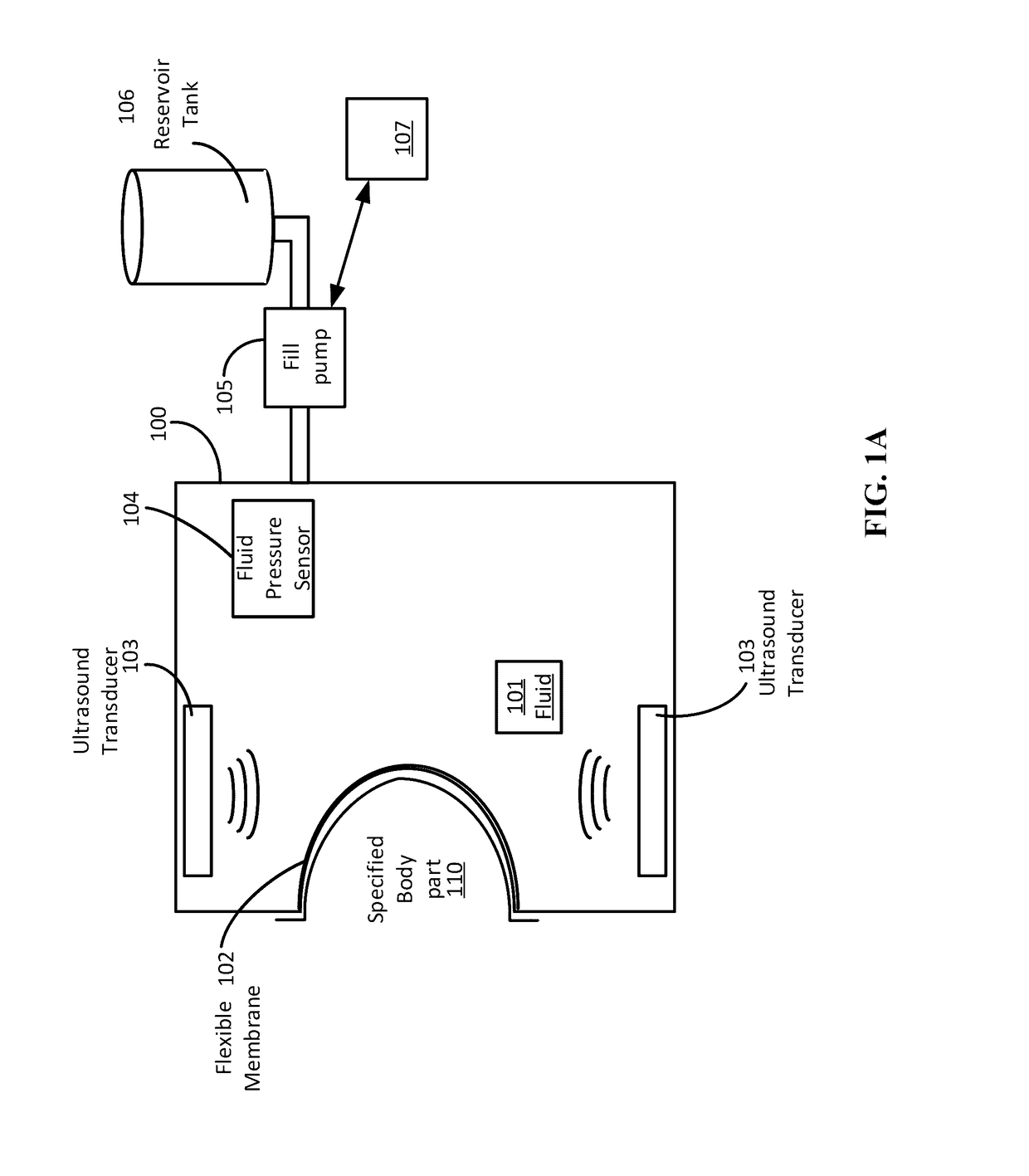
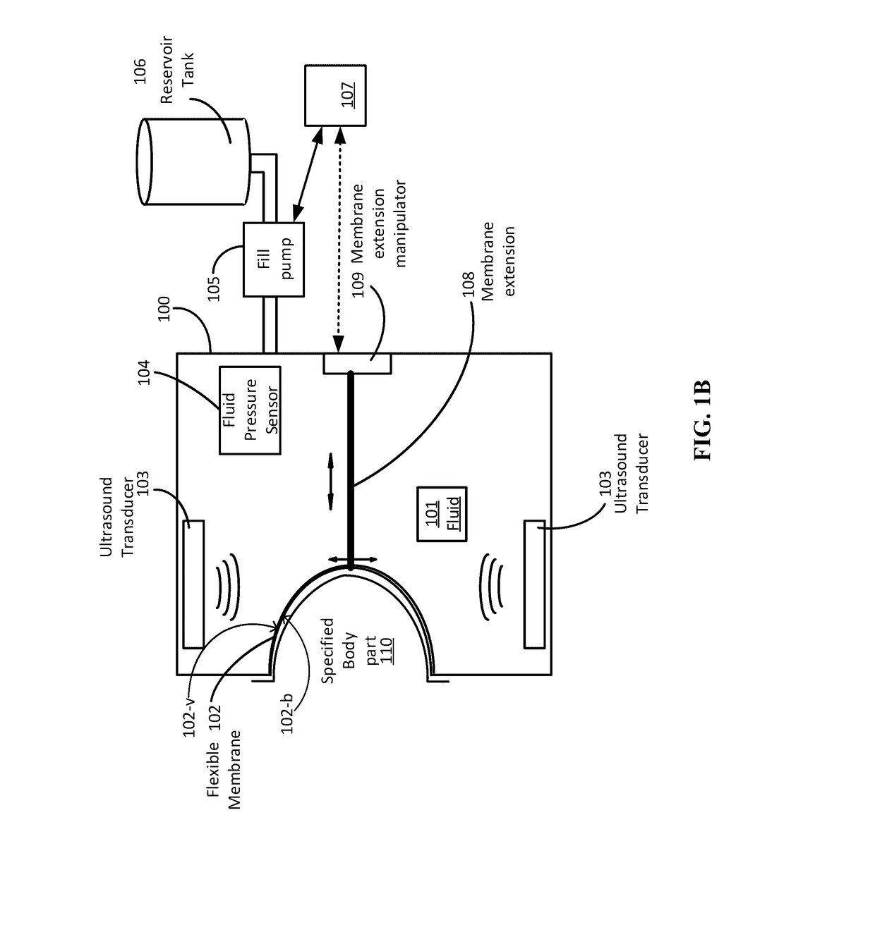
[0016]During a medical imaging procedure on a patient, the patient can rest their body part to be imaged against a flexible membrane that holds their body part in place using fluid in a vessel on the opposite side of the flexible membrane. Body parts that may be enclosed and retained in this manner by the flexible membrane include, but are not limited to, breast, hand, arm, leg, knee, shoulder, head, chest or the entire body. The arrangement and size of the membrane (and vessel) can be configured in a manner suitable for retention of a particular one or more body parts.

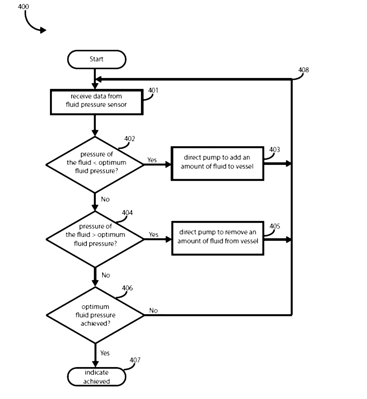
[0017]The comfort positioning in which the patient's body part is held in place can be accomplished by det...


PUM


Abstract

Systems and methods for the retention and stabilization of anatomy for ultrasound imaging are described. A system can include a vessel with a flexible membrane covering an opening, a fluid pressure sensor, a pump, and a pump controller for adding and removing fluid from the vessel until an optimum fluid pressure value or range of values is achieved. A method can include identifying an optimum fluid pressure range or value for retaining a specified body part in a stable position against a flexible membrane, monitoring the pressure of fluid in a vessel having the flexible membrane covering an opening, and directing a pump to add or remove fluid until the optimum fluid pressure value or range of values is achieved.

Description

BACKGROUND[0001]Patient motion is the single largest cause of image artifacts in medical imaging systems. As these medical imaging systems are developed to achieve higher and higher resolutions, even very small patient motions can blur the imaging results. Physically restraining the patient can be very uncomfortable for the patient. For sensitive body parts, this can be a particularly difficult issue as people will tend to postpone or simply not do important medical imaging screening tests like mammography to avoid patient discomfort. Improving patient comfort can improve patient compliance with necessary or recommended medical imaging procedures as well as the effectiveness of the medical imaging by reducing the occurrence of image artifacts.BRIEF SUMMARY[0002]Systems and methods for retention and stabilization of anatomy for ultrasound imaging are described. In some cases, body parts can be stabilized against a flexible membrane for patient comfort and minimizing imaging artifacts...

Claims


Application Information

Patent Timeline
Patent Type & Authority Applications(United States)
IPC IPC(8): A61B8/00A61B8/08
CPCA61B8/4209A61B8/0825A61B8/587A61B8/4281A61B8/403
Inventor LENOX, MARK WAYNEPIRSHAFIEY, NASSER CHARLESMALIK, BILAL HAMEEDKLOCK, JOHN CHARLES
Owner QT ULTRASOUND
Why Eureka
Try Eureka