Methods for tissue fabrication

Inactive Publication Date: 2017-05-11
ORGANOVO
0 Cites 11 Cited by

AI-Extracted Technical Summary

Problems solved by technology

One of the problems in tissue engineering is achieving and maintaining compartmentalization of cell types within a tissue.
Disadvantages of this method are that very high concentrations of divalent cross-...

Method used

[0032]An advantage of the engineered tissues and methodologies described herein is that they allow retention of the shape of the structure without compromising the functionality of the original cell types, and that they require no use of potentially toxic cross-linkers such as high ion levels, enzymes or UV light. The shape of the bioprinted structure is advantageously maintained by multiple approaches. A cold exposure step and/or an incubation step at below 37° C. are advantageous in that they allow for printed bio-inks to better maintain their shape during maturation, and limit the bioprinted cells exposure to cross-linkers that may damage the cells, such as supraphysiological levels of calcium, and reduces apoptosis in the resulting bioprinted tissues (FIG. 16). Surprisingly, this works with multiple tissue types, and even tissue types that are normally internal to the body, and, thus, at 37° C. (FIG. 11, kidney tissue). A temporal delay between printing bio-inks allows for maturation of a basal layer before application of any succeeding layer. The invention also incorporates a novel aerosol spray printing method into a 3D tissue model. The aerosol spray approach provides a for a unique discontinuous method compared to a continuous deposition method in that it allows the creation of a thinner layer, and allows for deposition of material onto an existing tissue layer after a period of maturation. This is advantageous because it may produce a tissue that better mimics native tissue in vivo. This can also be advantageous because it can reduce the number of cells required and allow for bioprinting with limited cell populations. This aerosol spray method can be applied to create multiple layers at multiple time points. For example, this method could be used for constructing a skin tissue by spraying first with undifferentiated keratinocytes followed by spraying with differentiated keratinocytes this could better mimic native skin.
[0045]One advantage to fabricating tissue with the bioprinting platform disclosed herein compared to current tissue models and natural tissue is that the process is automated. This allows for greater reproducibility and scalability. For example, it is possible to miniaturize the tissue geometry in order to print bio-ink into well plate formats such as 6, 12, 24, 48, 96, 384 or 1536-well plates for use in screening applications including high-throughput screening applications. Another major advantage of an automated platform is that it can be utilized to administer substances for toxicity testing in addition to bioprinting tissue. Current testing in tissue models is limited by the manual approaches necessary both to fabricate the tissue and to apply a test material to that tissue, limiting the application to topical administration. The flexibility of the printing platform allows for a variety of methods for application, deposition, and incorporation into tissues not possible with a manual approach. For example, test substances could be sprayed in a fine mist using the aerosol spray technology, or injected into the dermal layer utilizing the continuous deposition module. A third major advantage of bioprinting in a tissue toxicology model is the time frame in which a layered structure can be generated and tested. Bioprinting approaches can overlay sheets of cells simultaneously or with a delay to create multiple layers which can then be allowed to mature and differentiate for a defined period of time. The bioprinting platform allows for longitudinal studies not possible with manual approaches because test or therapeutic substances can be exposed to or incorporated into tissues during printing or administered to mature tissues at later time points.
[0054]In some embodiments, the engineered tissues, arrays, and methods described herein incorporate continuous deposition printing into a 3D tissue model. Continuous deposition is optionally utilized to produce single or multiple layers. In one embodiment, a bio-ink comprised of fibroblasts is printed to produce a tissue mimicking the dermis. In another embodiment, bio-ink comprised of keratinocytes or a mixture of keratinocytes and melanocytes is printed to produce a tissue to mimic the epidermis. A third embodiment combines bio-inks to simultaneously deposit the epidermal bio-ink on top of the dermal bio-ink. Continuous deposition printing provid...

Benefits of technology

[0002]While tissue engineering holds great potential for mankind, many problems must be overcome before the full extent of these advantages can be realized. One of the problems in tissue engineering is achieving and maintaining compartmentalization of cell types within a tissue. While bioprinting overcomes some of those challenges in the initial fabrication step, new methods are needed that are broadly applicable and support achievement and maintenance of cellular compartments post fabrication without compromising cell viability and function. For example, one way to induce compartmentalization is to utilize calcium-cross-linked hydrogels as a component of...

Abstract

Disclosed herein are improved methods for fabricating bioprinted, three-dimensional, biological tissues. The methods relate to exposures to low temperatures, incubations at low temperatures of various durations, and fabrication in environments without structural cross-linking treatments.

Application Domain

Artificial cell constructs3D culture

Technology Topic

Biological tissueBiology

Image

  • Methods for tissue fabrication
  • Methods for tissue fabrication
  • Methods for tissue fabrication

Examples

  • Experimental program(4)

Example

Example 1—Incubation Below 37° C. Improves Skin Tissue Formation
Procedures
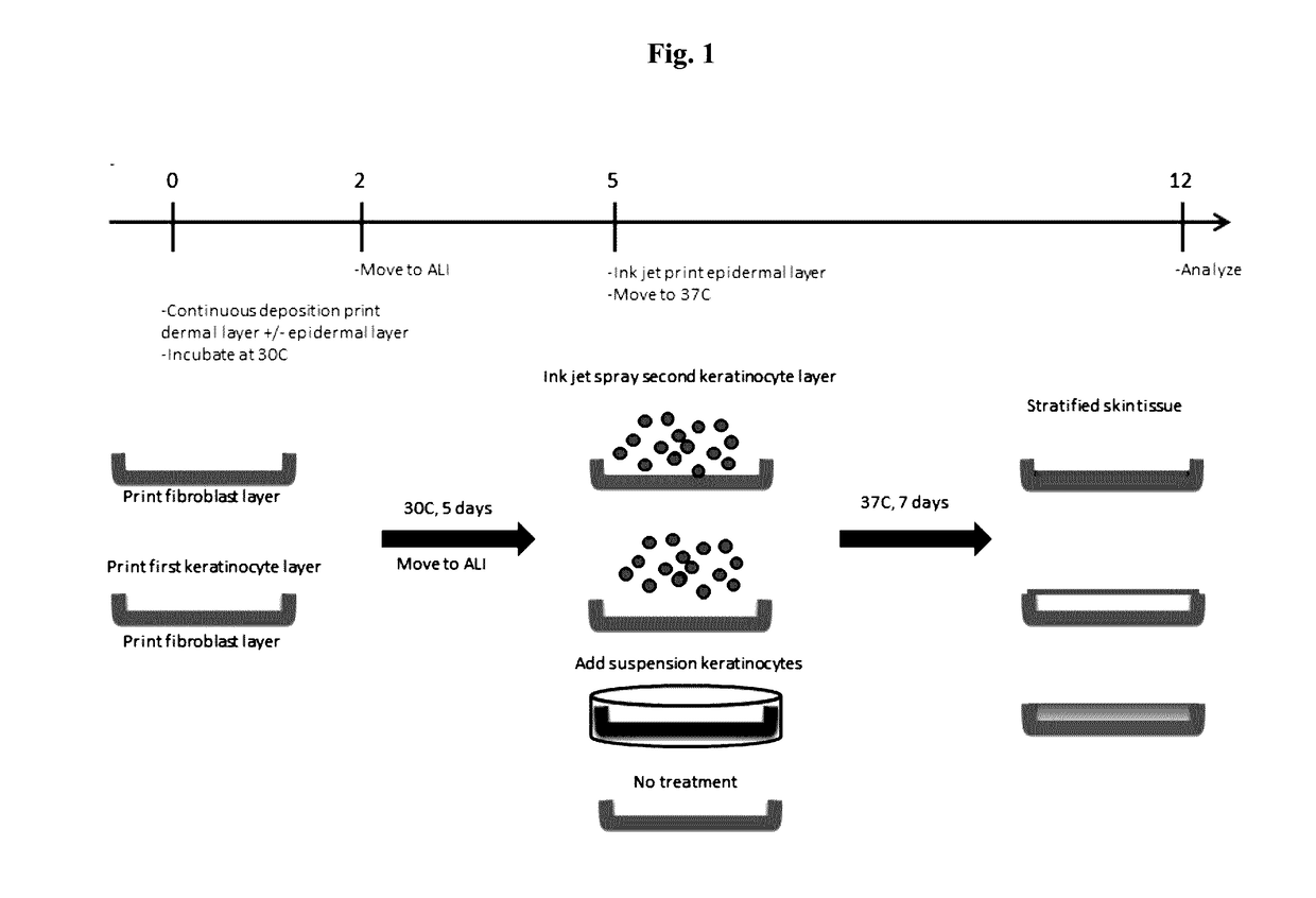
[0088]Bio-ink was generated by a cellular mixture of 100% primary adult human dermal fibroblasts (HDFa) in 6% gelatin (Novogel® 2.0) in a concentration of 150 million cells per milliliter.
[0089]Three-dimensional bio-ink constructs were printed by continuous deposition using the Novogen Bioprinter® platform in a 4 mm×4 mm×0.5 mm base sheet with a 1 mm wall bordering the top to create a dermal structure resembling a cup. One tissue construct was printed per transwell in a 6 well plate. The transwell printing surface contained a polytetrafluoroethylene (PTFE) membrane coated with equimolar mixture of types I and III collagen (bovine) with pores 3 μm in size.
[0090]Epidermal cell paste containing a mixture of 95% primary adult human epidermal keratinocytes (HEKa) and 5% primary adult human epidermal melanocytes (HEMa) was then printed on top of the dermal bio-ink immediately or between 0.020 seconds and several hours or several days.
[0091]Cell paste was measured post print at 90.5% viable by trypan exclusion assay. Cell number in deposited epidermal layer was estimated at 160,000 cells by cell counting on a Cell-O-Meter.
[0092]Media was then added to the outer well of the transwell in a volume of 2 ml. The media used for subsequent growth and maintenance of the skin tissue was a 50:40:10 ratio of HDFa:HEKa:HEMa media. The volume added was sufficient to collect at the base of the printed structure but not to submerge the structure. Media was changed 48 hours later and subsequently changed daily after that.
[0093]Printed constructs were placed into a non-humidified incubator at 30° C. for 5 days. This is a key step that enables maintenance of tissue shape for a period of maturation. After 5 days, the temperature was raised to 37° C. for an additional 7 days.
[0094]At days 2, 9, and 12, constructs were either lysed for RNA analysis or fixed in 2% PFA for histological analysis.
Results
[0095]H&E staining of skin tissues at day 12 shows a distinct layered architecture (FIGS. 1 and 3). Fibroblasts in a dermal layer are observed at the base (purple) and differentiated keratinocytes in an epidermal layer (pink) on top. An unexpected finding with this approach is the extent of the layered architecture observed. In particular, there is a layer of cells with distinct morphology can be observed at the interface (arrows). This layer stains specifically for CK14, indicating that the keratinocyte cells in the deposited paste have arranged into a basal layer.
[0096]Distinct layers of differentiated keratinocytes are visualized by simultaneously staining for a basal cell marker CK5 and involucrin (IVL), a later stage differentiation marker of granular and cornified keratinocytes. Similar to normal human skin, differences in morphology are seen as basal cells appear to have a distinct cuboidal morphology, while differentiated keratinocytes on top appear flatter. The layered architecture also includes CK10-positive spinous and granular keratinocytes in mid stages differentiation (FIG. 4).
[0097]Although previous print methods have resulted in CK14 positive staining of the epidermal layer, the observed pattern is widespread throughout the layer and non-specific to a basal region at day 10. In the current approach, what is unexpected is that the staining is limited to a defined region at the base of the epidermal layer similar to native human skin at day 12 (FIG. 5).
[0098]Gene expression analysis supports histological findings. Data shows an increase in epidermal differentiation markers CK1, CK10, and especially late marker FLG over time. Gene expression also shows that collagen 4 levels increase over time, suggesting formation of a basement membrane. Collagen I levels are maintained over the time course of the experiment suggesting dermal layer remains viable (FIG. 6).

Example

Example 2—Transient Exposure at 4° C. and Incubation Below 37° C. Improves Skin Tissue Formation
Procedures
[0099]Bio-ink was generated by a cellular mixture of 100% primary adult human dermal fibroblasts (HDFa) in 8% gelatin (Novogel®) in a concentration of 100 million cells per milliliter. The cell:gelatin ratio was altered to reduce the cellular density of the dermal sheet to better mimic dermal tissue in native skin.
[0100]Three-dimensional bio-ink constructs were printed by continuous deposition using the Novogen Bioprinter® platform in a 4 mm×4 mm×0.5 mm base sheet to create a dermal structure resembling a sheet. One tissue construct was printed per transwell—in a 6 well plate. The transwell printing surface contained a polytetrafluoroethylene (PTFE)-membrane coated with equimolar mixture of types I and III collagen (bovine) with pores 3 μm in-size.
[0101]Epidermal cell paste containing a mixture of 100% primary neonatal human epidermal keratinocytes (HEKn) was then printed on top of the dermal bio-ink. A separate but identical epidermal paste structure was simultaneously deposited next to the dermal sheet directly onto the transwell printing surface. This structure was only comprised of epidermal keratinocyte paste and contained no dermal tissue.
[0102]Cell paste was measured post print at 87.1% viable by trypan exclusion assay. Cell number in deposited epidermal layer was estimated at 60,000 cells by cell counting on a Cell-O-Meter. Immediately following the print, constructs were placed in 4° C. for 10 minutes. This is a key step to harden the Novogel®, which helps to maintain the printed shape and improve construct to construct uniformity.
[0103]Cold media was then added to the outer well of the transwell in a volume of 3 ml. The media used for subsequent growth and maintenance of the skin tissue was a 50:50 ratio of HDFa:HEKn media. The initial volume added was sufficient to submerge the structure. All subsequent media changes used warmed media (30-37° C.) added to the outer well of the transwell and not to the inner basket. Media was changed 48 hours later and reduced to a volume of 1.5 ml per well to bring the structure to an air-liquid interface (ALI). Media and subsequently changed 48 hours after that (day 4) at a volume of 1.5 ml. On day 5, media was changed and further reduced to 1 ml per well and subsequently changed daily
[0104]Printed constructs were placed into a non-humidified incubator at 30° C. for 5 days. This is a key step that enables maintenance of tissue shape for a period of maturation. After 5 days, the temperature was raised to 37° C. for an additional 7 days.
[0105]At days 0 and 12, constructs were either lysed for RNA analysis or fixed in 2% PFA for histological analysis.
Results
[0106]Subsequent histological analysis to compare epidermal layer patterning of paste that had been printed on top of a dermal sheet versus directly onto the transwell surface yielded unexpected findings (FIGS. 7A and B). H&E staining of skin tissues at day 12 shows a distinct layered architecture only in structures with epidermal paste printed on top of a dermal layer (FIGS. 7 C and D versus F and G, FIG. 8A). Fibroblasts in a dermal layer are observed at the base (purple) and differentiated keratinocytes in an epidermal layer (pink) on top. In particular, there is a layer of cells with distinct morphology that can be observed at the interface. Distinct layers of differentiated keratinocytes are visualized by simultaneously staining for a basal cell marker CK5 (green) and involucrin (IVL, red), a later stage differentiation marker of granular and cornified keratinocytes (FIG. 7E versus H, FIG. 8B). The distinct green layer indicates that the keratinocyte cells in the deposited paste have arranged into a basal layer with a layer of more differentiated IVL positive cells on top. Similar to normal human skin, differences in morphology are seen as basal cells that appear to have a distinct cuboidal morphology, while differentiated keratinocytes on top appear flatter.
[0107]Staining for the proliferation marker PCNA (FIG. 8E, green) indicates that proliferation is high in both dermal fibroblasts and basal layer keratinocytes but not in differentiating keratinocytes. This pattern is similar to that which is found in native skin. Staining for apoptosis by TUNEL (FIG. 8F) also low showing very few positive staining cells in either dermal or epidermal layer. Collectively PCNA and TUNEL staining demonstrate that both dermal and epidermal compartments of the full thickness tissue are viable at day 12.
[0108]Gene expression analysis supports histological findings. Data shows an increase in mid epidermal differentiation markers CK1, CK10, and later markers IVL, Loricrin, and at day 12 compared to day 0. Gene expression also shows that collagen I and 4 levels are maintained over the time course of the experiment, while collagen 3 levels increase suggesting the dermal layer remains viable and functional (FIG. 9).
[0109]A number of surprising results were determined from this; for example, that epidermal paste can stratify into a distinct layered architecture. Current 3D skin models rely on differentiation of a single keratinocyte monolayer over an extended period of time to achieve this. Here we show that stratification is possible to achieve with a paste. The thickness of the paste is greater than a monolayer where the monolayer is approximately 18-20 microns) and shows that cells can self-organize within the paste and differentiate as layers. Also, we show that the keratinocyte paste printed directly onto the transwell surface without the presence of dermal tissue did not organize into stratified layers. Staining for the same differentiation markers shows mixed expression with no defined layers or distinct cell morphology. This unexpected finding indicates that the dermal layer directs differentiation and/or stratification of the epidermal keratinocytes, and that there is a uniqueness to the combination of dermal and epidermal cells that is not present in the epidermal cells alone. 3) The extent of the layered architecture observed in the tissues comprised of both epidermal and dermal cells including the staining of the CK5-positive basal layer which is limited to a defined region at the base of the epidermal layer similar to native human skin. The layered architecture also includes a CK10 positive (FIG. 8C) spinous and granular keratinocytes in mid stages differentiation and with a morphologically distinct cornified layer of keratinocytes visible by H&E and Trichrome staining above that (FIGS. 8A and D respectively).
[0110]A noteworthy advantage to this approach is the appearance of the dermal layer. H&E staining shows that the dermal fibroblasts do not form a thin sheet as in earlier examples 1 and 2, but a thicker structure. Collagen deposition, which is a key indicator of normal fibroblast function in the dermis, can be seen by both trichrome staining (blue color) and by immunofluorescent staining for collagen 3 (red) in between dermal cells (FIG. 8).

Example

Example 3—Incubation Below 37° C. Improves Kidney Tissue Formation
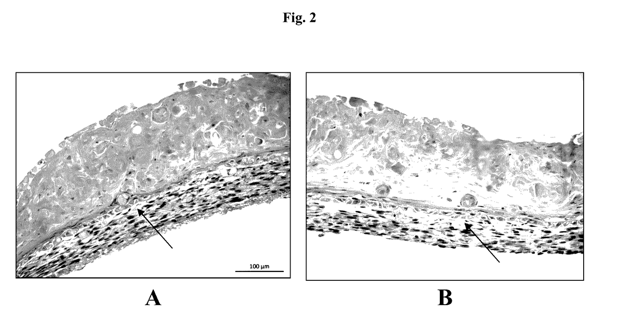
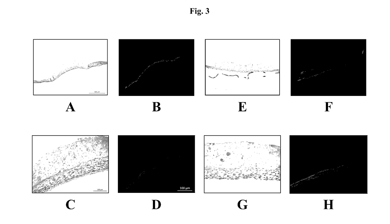
[0111]The interstitial layer of the renal proximal tubule model is composed of renal fibroblasts and HUVECs in Novogel®. To reduce the thickness and cellularity of the interstitial layer, the cell ratio was changed to 50% fibroblasts/50% HUVEC the concentration of the cells was 125 million cells/mL. Attempts to fabricate tissues using these cell ratios were hampered by a propensity of the tissues to “ball up,” preventing the sort of thin, spread out interstitial layer that is ideal. To assist in maintenance of construct shape following bioprinting, tissues were incubated at 30° C. for 3 days following printing to slow the rate of Novogel® dissipation.
[0112]The results show a tissue that better retains its overall dimensions (FIGS. 11C and D) after 30° C. incubation, when compared to 37° C. incubation (FIGS. 11A and B), and allows the cells to proliferate and secrete ECM to replace the Novogel® material as a binding agent (FIG. 11B). Following this maturation step, tissues can be shifted to 37° C. for the addition of epithelial cells to the structure, with no loss of tissue features or viability as a result of the culture time at 30° C. H&E staining is shown (FIG. 12A), and brush borders are indicated (FIG. 13 A, arrows). Trichrome staining indicates collagen secretion (FIGS. 12B, blue and 13B, arrows), and CD31 staining indicates the presence of HUVEC networks (FIG. 14, asterisks). Bioprinted tissues demonstrated γ-glutamyl-transferase activity which increases over time in culture, which is indicative of a functioning epithelial layer (FIG. 15). Considering that the kidney is a fully internal structure normally kept at 37° C., it is unexpected that the culture time at 30° C. would result in a tissue with the desired structural and functional characteristics.

PUM

no PUM

Description & Claims & Application Information

We can also present the details of the Description, Claims and Application information to help users get a comprehensive understanding of the technical details of the patent, such as background art, summary of invention, brief description of drawings, description of embodiments, and other original content. On the other hand, users can also determine the specific scope of protection of the technology through the list of claims; as well as understand the changes in the life cycle of the technology with the presentation of the patent timeline. Login to view more.
Why Eureka
Try Eureka