Microfluidic Processing of Leukocytes for Molecular Diagnostic Testing
a technology of microfluidic processing and leukocytes, applied in the field of microfluidic processing of leukocytes for molecular diagnostic testing, can solve problems such as loss and damage of cells, and achieve the effects of improving cell quality, reducing costs, and improving quality
- Summary
- Abstract
- Description
- Claims
- Application Information
AI Technical Summary
Benefits of technology
Problems solved by technology
Method used
Image
Examples
example 1
Fabrication
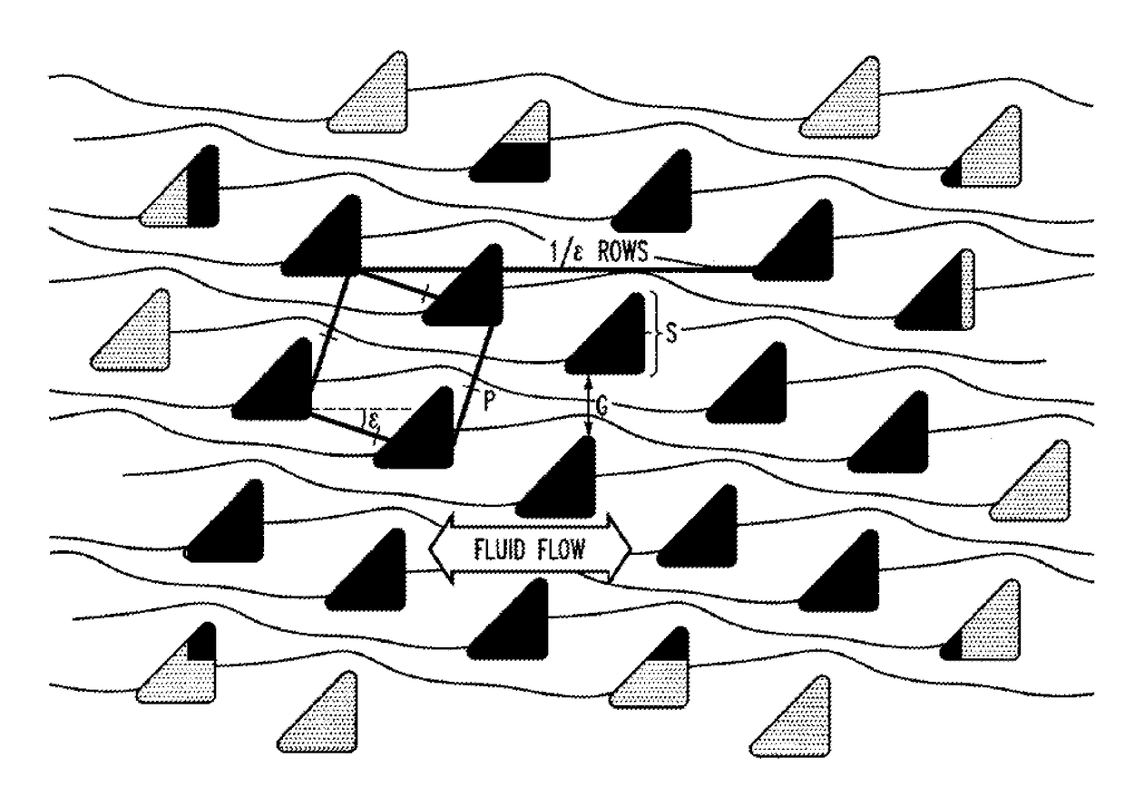
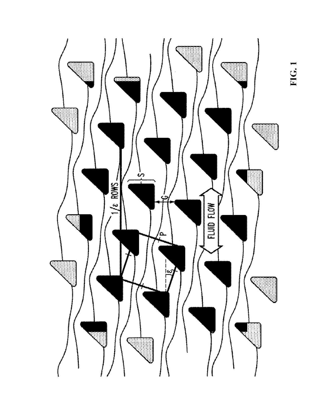
[0203]Chips are fabricated using highly anisotropic deep reactive ion etching (DRIE) in crystalline silicon polished substrates using a “Bosch” process which cycles between etching and sidewall passivation steps, so the post sidewall differs from vertical by only ˜1° . Optical lithography defines the patterns. Through-holes are micro-machined through the substrate enable fluid loading / unloading from the backside, which are mated to a plastic jig with connectors to input sources and output collection. The chip is pre-treated with Triblock copolymer F108 (2g / 1) to reduce cell adhesion. The chip design parameters (e.g. critical size for bumping behavior) are adjusted to obtain a high yield.
example 2
Operation
[0204]Leukocytes from 0.1-1 ml of erythrocyte-lysed whole blood (optionally diluted with buffer (PBS without calcium and magnesium, containing 1% BSA and 4mM EDTA), and optionally spiked with leukemia cells) are incubated (“immunostained”) with fluorescent Mabs against multiple leukocyte differentiation cell surface antigens (i.e. CD45 / CD14 / 15 (to enumerate monogranulocytic cell types), CD3 / 4 / 8 (to enumerate the common T lymphocyte subsets), CD19 / 56 / 14 (to identify B lymphocytes and NK cells), CD45 / CD235a / CD71 (to identify any contaminating erythroid cells) and with a viability dye. This is done conventionally, i.e. off chip. Cells are then washed and concentrated to ˜1-10 million cells / ml using DLD chips designed to move leukocytes and leukemia cells from the initial stream of the input cell suspension containing fluorescent Mabs to the output stream of fresh buffer against the chip wall (FIG. 17B).
[0205]The method can recover >90% of the input leukocytes, concentrated bac...
example 3
Leukocytes from UCB
[0208]Leukocytes can be harvested from a variety of tissues. Table 2 shows leukocyte enrichment experiments from umbilical cord blood (UCB). The starting sample is 3 ml UCB, diluted 1:1 with running buffer. The leukocyte-enriched output product contained erythrocyte levels below detection (Hemavet cell counter), so product purity is determined by multicolor FACS analysis using labels against CD45, CD14, CD235a, and a viable nucleic acid dye. For the combined fractions, erythrocyte depletion is 99%, leukocyte recovery is 87%, and leukocyte purity (i.e. 100%-% erythrocytes) is 81-88%. There is some dead volume the instrument configuration, so a small portion of sample remains in the system and is not processed. With some minor engineering changes, the full sample can be sorted, and the leukocyte recovery may rise to ∃90%. Viability by trypan blue dye exclusion is >90% in all fractions. Granulocytes, lymphocytes, and monocytes are close to the initial “differential l...
PUM
| Property | Measurement | Unit |
|---|---|---|
| volume | aaaaa | aaaaa |
| tilt angle | aaaaa | aaaaa |
| tilt angle | aaaaa | aaaaa |
Abstract
Description
Claims
Application Information
 - R&D
- Intellectual Property
- Life Sciences
- Materials
- Tech Scout
- Unparalleled Data Quality
- Higher Quality Content
- 60% Fewer Hallucinations
Browse by: Latest US Patents, China's latest patents, Technical Efficacy Thesaurus, Application Domain, Technology Topic, Popular Technical Reports.
© 2025 PatSnap. All rights reserved.Legal|Privacy policy|Modern Slavery Act Transparency Statement|Sitemap|About US| Contact US: help@patsnap.com


