

Personal brain structure displaying device having intracranial electrodes and its displaying method
a brain structure and display device technology, applied in diagnostic recording/measuring, tomography, application, etc., can solve the problems of the inability to apply brain function maps (or brain atlases), and achieve poor recording or stimulation effect of electrodes
- Summary
- Abstract
- Description
- Claims
- Application Information
AI Technical Summary
Benefits of technology
Problems solved by technology
Method used
Image

Examples
Embodiment Construction
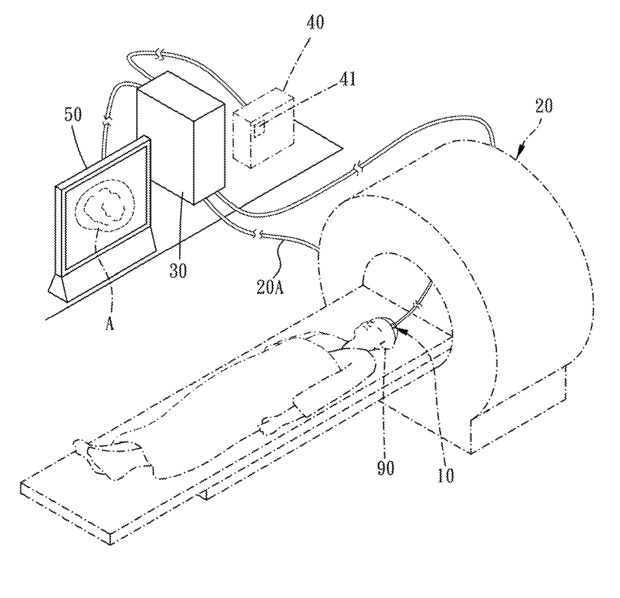
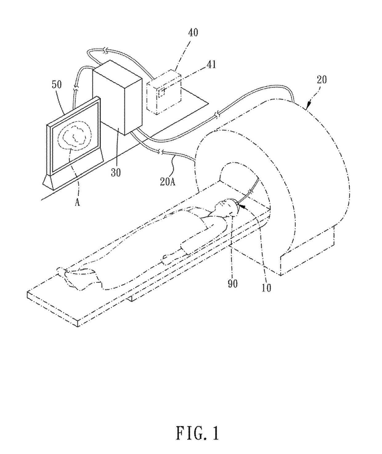
[0043]Referring to FIGS. 1, 2, and 3, this invention relates to a personal brain structure displaying device having intracranial electrodes and its displaying method. The device of this invention, it mainly includes an electrode module 10, an image capturing module 20, a controller 30, a brain functional map adjusting portion 40, and a displaying portion 50.
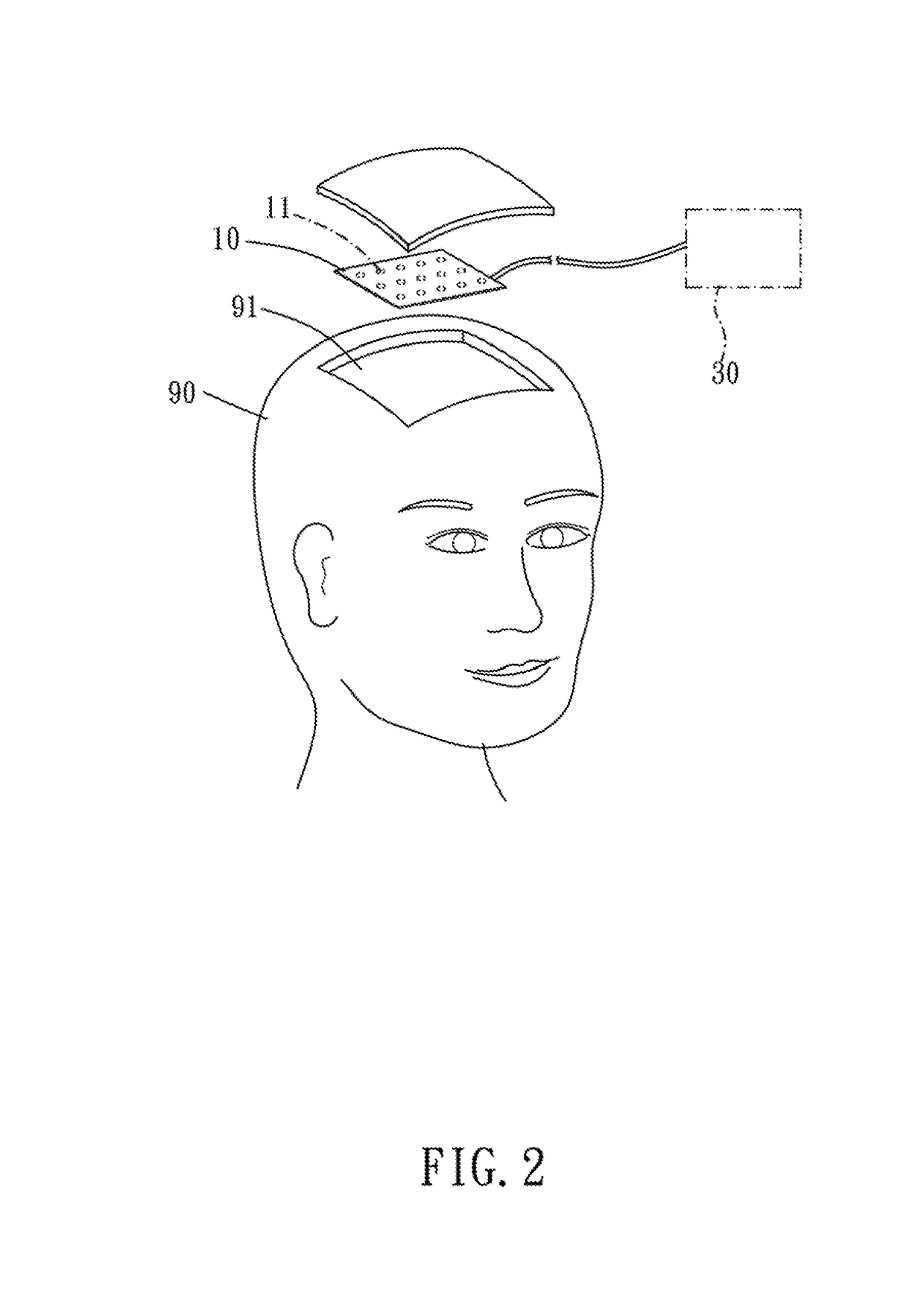
[0044]With regard to this electrode module 10, it is positioned inside an intracranial portion 91 of a human head 90. The electrode module 10 has multiple electrodes 11.
[0045]About the image capturing module 20, it is provided for capturing a brain area image (referring to FIG. 4) of the human head 90. This image capturing module 20 is able to obtain a three-dimensional (3D) brain information 20A which includes a plurality of two dimensional (2D) cross-sectional images 21 (illustrated as FIGS. 5A, 5B, and 5C; having a first image width D1, a second image width D2, and a third image width D3 respectively). Each 2D cross-sectional ...
PUM
 Abstract
Description
Claims
Application Information