A method and device for fusion display of cerebral cortex electrodes and magnetic resonance images

A magnetic resonance image fusion display technology, applied in the field of information fusion, can solve the problem of unfavorable acquisition of electrode position information

Active Publication Date: 2019-01-18
SHENZHEN SINORAD MEDICAL ELECTRONICS
View PDF4 Cites 0 Cited by
  • Summary
  • Abstract
  • Description
  • Claims
  • Application Information

AI Technical Summary

Problems solved by technology

[0004] The technical problem to be solved by the present invention is to provide a method and device for fusion display of cerebral cortex electrodes and magnetic resonance images that can obtain the position information of the electrodes in view of the above-mentioned defects in the prior art that are not conducive to obtaining the position information of the electrodes

Method used


Image

  • A method and device for fusion display of cerebral cortex electrodes and magnetic resonance images
  • A method and device for fusion display of cerebral cortex electrodes and magnetic resonance images
  • A method and device for fusion display of cerebral cortex electrodes and magnetic resonance images

Examples


Embodiment Construction

[0083] The following will clearly and completely describe the technical solutions in the embodiments of the present invention with reference to the accompanying drawings in the embodiments of the present invention. Obviously, the described embodiments are only some of the embodiments of the present invention, not all of them. Based on the embodiments of the present invention, all other embodiments obtained by persons of ordinary skill in the art without creative efforts fall within the protection scope of the present invention.

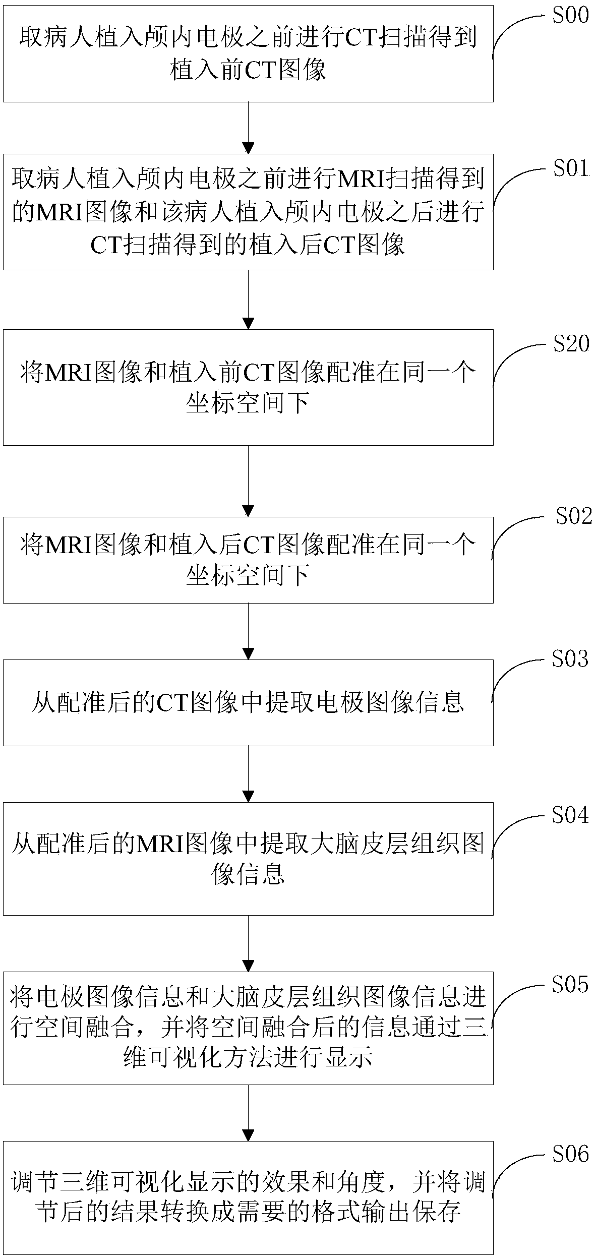
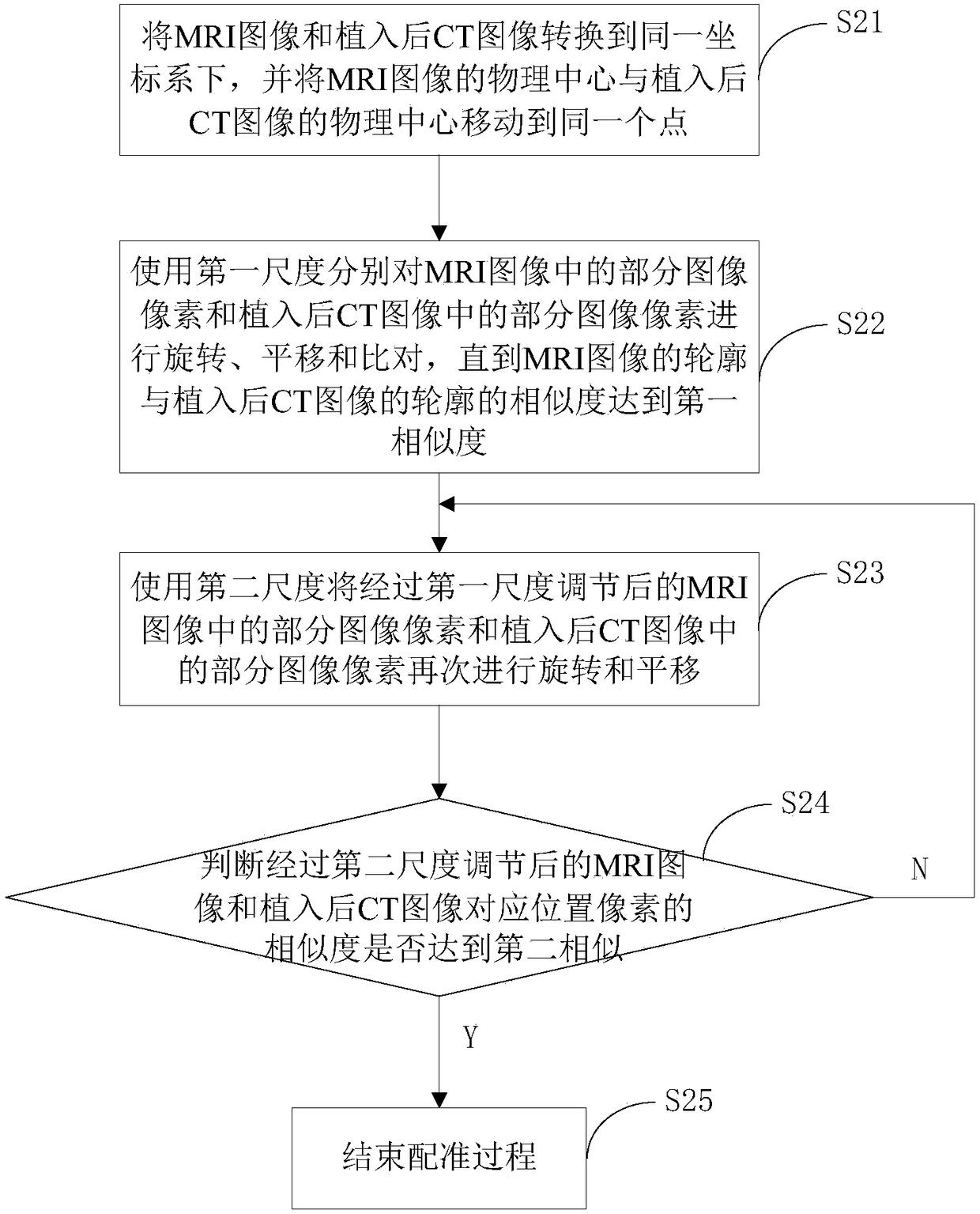
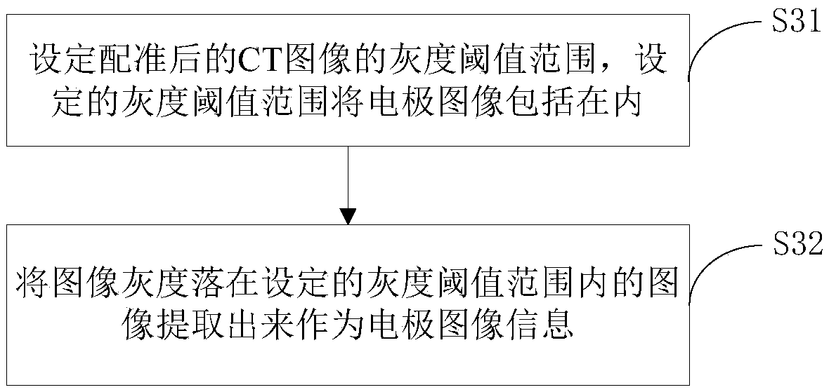
[0084] In the embodiment of the method and device for fusion display of cerebral cortex electrodes and magnetic resonance images of the present invention, the flow chart of the method for fusion display of cerebral cortex electrodes and magnetic resonance images is as follows figure 1 shown. figure 1 Among them, the method for fusion display of cerebral cortex electrodes and magnetic resonance images includes the following steps:

[0085] Step S01 ta...


PUM


Abstract

A method and an apparatus for fusion display of cerebral cortex electrode and magnetic resonance images. The method comprises: extracting an MRI image obtained by MRI scanning before an intracranial electrode is implanted into a patient and a post-implantation CT image obtained by CT scanning after the intracranial electrode is implanted into the patient (S01); registering the MRI image and the post-implantation CT image under an identical coordinate space (S02); extracting electrode image information from the registered CT image (S03); extracting cerebral cortex tissue image information from the registered MRI image (S04); spatially fusing the electrode image information and the cerebral cortex tissue image information, and displaying the spatially-fused information by means of a three-dimensional visual method (S05); and adjusting a three-dimensional visual display effect and angle, converting an adjustment result into a needed format for output and storage (S06). By implementing method and apparatus for fusion display of cerebral cortex electrode and magnetic resonance images, position information about electrodes can be acquired.

Description

technical field [0001] The invention relates to the field of information fusion, in particular to a method and device for fusing and displaying cerebral cortex electrodes and magnetic resonance images. Background technique [0002] Surgery is an important means of treating certain brain diseases. The key to surgical treatment is to locate the lesion, that is, to find out the abnormally excited neuron group. At present, intracranial electrodes are considered to be the gold standard for locating lesions. Intracranial electrodes can directly record the EEG activity of the cerebral cortex and internal brain tissue, and can detect abnormal cortical discharges within 1 cm. It has the advantages of clear graphics and high sensitivity. For those cases where the lesion cannot be clearly identified by non-invasive examination, EEG monitoring with intracranial electrodes is a safe and reliable positioning method. [0003] After the electrodes are implanted in the brain, information o...

Claims


Application Information

Patent Timeline
Patent Type & Authority Patents(China)
IPC IPC(8): G06T7/00G06T3/00
CPCG06T3/0068G06T7/0012G06T2207/10081G06T2207/10088G06T2207/20221G06T2207/30016G06T2207/30088
Inventor 汤洁李卓任乐枫胡楠谢菲
Owner SHENZHEN SINORAD MEDICAL ELECTRONICS
Why Eureka
Try Eureka