Multimodal silica-based nanoparticles

a technology of nanoparticles and fluorescence, which is applied in the field of fluorescence silica-based nanoparticles, can solve the problems of limited imaging technology, disproportionate number of invasive biopsies, and limited evaluation of metastatic disease spread and tumor margins, especially in areas of complex anatomy

Active Publication Date: 2018-04-05
CORNELL UNIVERSITY +2
View PDF0 Cites 5 Cited by
  • Summary
  • Abstract
  • Description
  • Claims
  • Application Information

AI Technical Summary

Benefits of technology

[0022]The present invention further provides a method for targeting a tumor cell comprising administering to a cancer patient an effective amount of a fluorescent silica based nanoparticle comprising a silica-based core comprising a fluorescent compound positioned within the silica-based core; a silica shell surrounding at least a portion of the core; an organic polymer attached to the nanoparticle; a ligand attached to the nanoparticle and capable of binding a tumor marker; and at least one therapeutic agent. The nanoparticle may be radiolabeled. The nanoparticle may be administered to the patient by, but not restricted to, the following routes: oral, intravenous, nasal, subcutaneous, local, intramuscular or transdermal.

Problems solved by technology

However, in surgical settings, the evaluation of metastatic disease spread and tumor margins, particularly in areas of complex anatomy, is limited by a lack of imaging technologies.
This has led to a disproportionate number of invasive biopsies.
Single fluorophores are usually not stable and have limited brightness for imaging.
Q-dots are generally made from heavy metal ions such as Pb2+ or Cd2+ and, therefore, are toxic.
A shortcoming of existing fluorescent nanoparticles is their limited brightness and their low detectability as fluorescent probes in dispersed systems.

Method used


Image

  • Multimodal silica-based nanoparticles
  • Multimodal silica-based nanoparticles
  • Multimodal silica-based nanoparticles

Examples


example 1 preparation

and Characterization of PEG-Coated Nanoparticles

[0218]Nanoparticles containing an NIR-emitting dye (Cy-5) were synthesized and functionalized by PEGylation according to well-established protocols as disclosed in PCT / US2008 / 074894 and Stober et al. Controlled growth of monodispersed silica spheres in the micron size range. Colloid Interface Sci. 1968; 26:62-69. Ohnishi et al. J. Mol. Imaging 2005, 4:172-181. Cy5 malemide was reacted with a co-reactive organo silane compound, (3-Mercaptopropyl)tromethoxysilane to form a fluorescent silica precursor. This fluorescent silica precursor was co-condensed with tetraethylorthosilicate to form a fluorescent silica based core. A PEG-silane compound, with methoxy-terminated poly(ethylene glycol) chains (PEG, ˜0.5 kDa) Methoxy(Polyethy leneoxy) Propyl]—Trimethoxysilane, was added to the fluorescent silica based core to form a PEG coating on the core. PEG-coated nanoparticles were dialyzed to physiological saline (0.15M NaCl in H2O) through 3500 ...

example 2

Renal Clearance of PEG Coated Nanoparticles

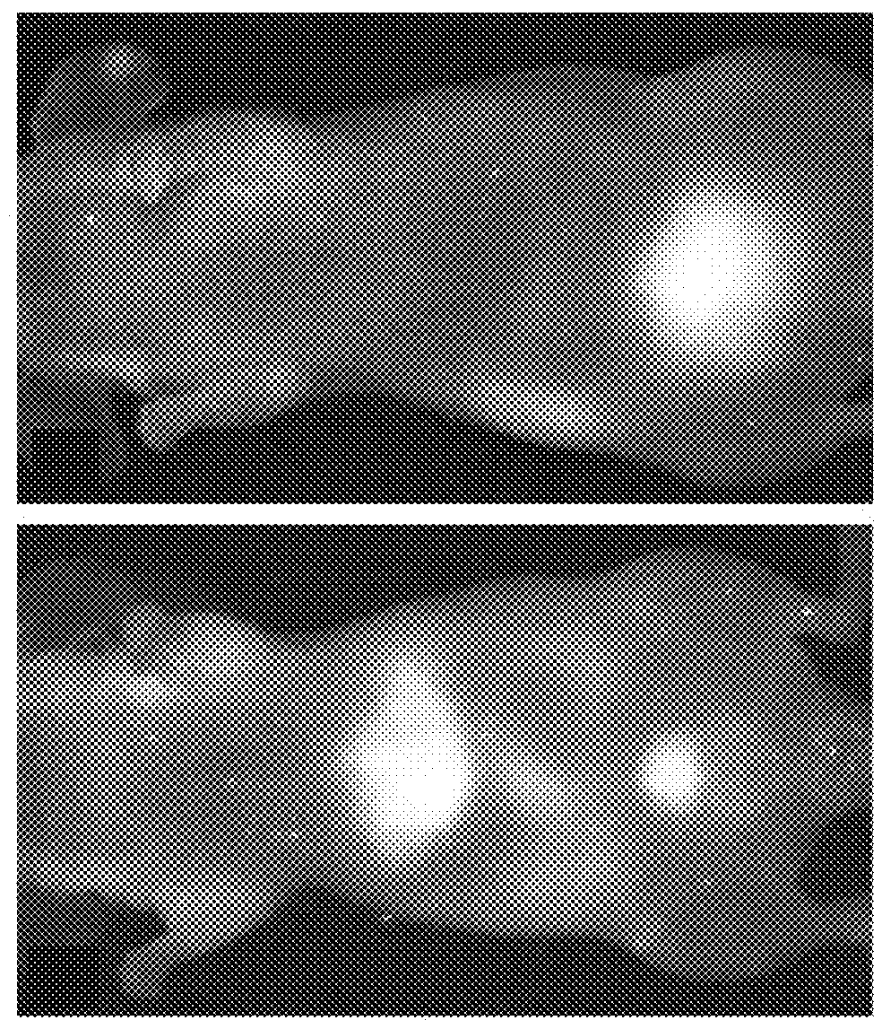
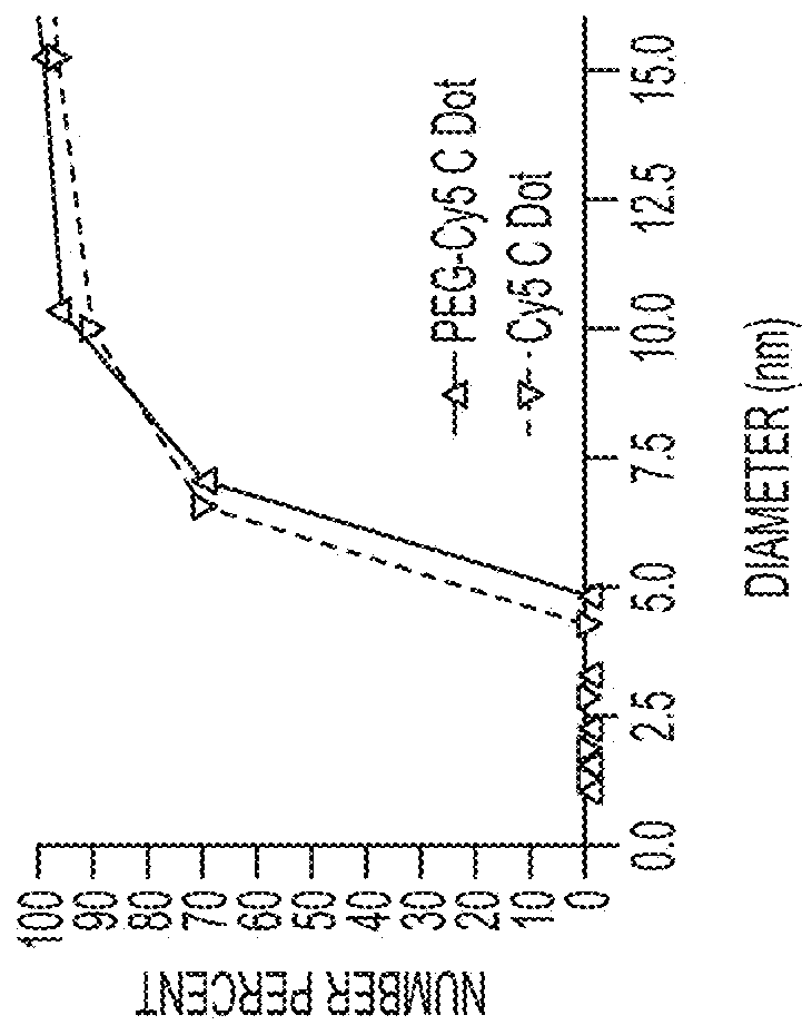
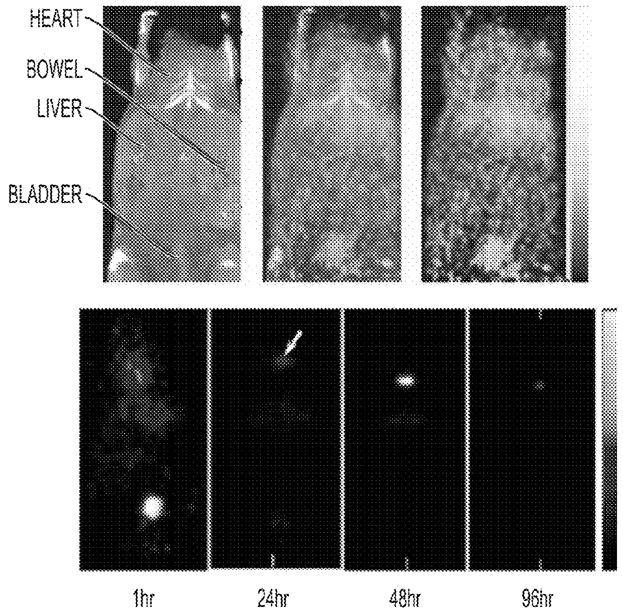
[0219]Fluorescent core-shell silica nanoparticles, having a hydrodynamic radius of about 3 nm, were synthesized. These nanoparticles were found to be in the 6-10 nm diameter range, as shown by dynamic light scattering (DLS) results (FIG. 1A). In vivo whole-body NIR fluorescence imaging of bare (no PEG coat) silica nanoparticles, on the order of 6-nm and 3.3-nm, in nude mice showed considerable renal clearance 45 min post-injection with a significant accumulation remaining in the liver (FIG. 1B). Eventual excretion into the enterohepatic circulation occurred during the ensuing 24 h. On the basis of these results, particles were covalently coated with methoxy-terminated poly(ethylene glycol) chains (PEG, ˜0.5 kDa), per protocols in PCT / US2008 / 074894, to prevent opsonization and further enhance particle clearance while maintaining a small hydrodynamic size. This treatment decreased liver retention and resulted in increased renal filtration int...

example 6

ping in Miniswine

[0270]Real-time intraoperative scanning of the nodal basin cannot be practically achieved at the present time, as these systems are generally too cumbersome and expensive for use in the operating suite or may be unable to provide the necessary field-of-view or tissue contrast. Further, there are no clinically promising, biostable fluorophore-containing agents, offering improved photophysical features and longer circulation lifetimes over parent dyes, available to enhance tissue contrast for extended nodal mapping / resection procedures. With this animal study, we will show that advances in both multimodal particle probes and real-time molecular imaging device technologies can be readily translated to a variety of future human clinical trials. Such transformative technologies can significantly impact standard intraoperative cancer care by providing state-of-the-art targeted visualization tools for facilitating metastatic SLN detection and enabling accurate delineation ...


PUM

PropertyMeasurementUnit
hydrodynamic diameteraaaaaaaaaa
diameteraaaaaaaaaa
diameteraaaaaaaaaa

Abstract

The present invention provides a fluorescent silica-based nanoparticle that allows for precise detection, characterization, monitoring and treatment of a disease such as cancer. The nanoparticle has a range of diameters including between about 0.1 nm and about 100 nm, between about 0.5 nm and about 50 nm, between about 1 nm and about 25 nm, between about 1 nm and about 15 nm, or between about 1 nm and about 8 nm. The nanoparticle has a fluorescent compound positioned within the nanoparticle, and has greater brightness and fluorescent quantum yield than the free fluorescent compound. The nanoparticle also exhibits high biostability and biocompatibility. To facilitate efficient urinary excretion of the nanoparticle, it may be coated with an organic polymer, such as poly(ethylene glycol) (PEG). The small size of the nanoparticle, the silica base and the organic polymer coating minimizes the toxicity of the nanoparticle when administered in vivo. In order to target a specific cell type, the nanoparticle may further be conjugated to a ligand, which is capable of binding to a cellular component associated with the specific cell type, such as a tumor marker. In one embodiment, a therapeutic agent may be attached to the nanoparticle. To permit the nanoparticle to be detectable by not only optical fluorescence imaging, but also other imaging techniques, such as positron emission tomography (PET), single photon emission computed tomography (SPECT), computerized tomography (CT), bioluminescence imaging, and magnetic resonance imaging (MRI), radionuclides / radiometals or paramagnetic ions may be conjugated to the nanoparticle.

Description

CROSS REFERENCE TO RELATED APPLICATIONS[0001]This application is a continuation of U.S. application Ser. No. 14 / 215,879, filed Mar. 17, 2014, which claims priority to U.S. Provisional Application No. 61 / 794,414, filed Mar. 15, 2013. The disclosures of each of the above-referenced applications are incorporated by referenced herein in their entireties.STATEMENT REGARDING FEDERALLY SPONSORED RESEARCH OR DEVELOPMENT[0002]This invention was made with government support under grant numbers CA086438, CA083084, CA008748, RR024996, and CA161280 awarded by National Institutes of Health. The government has certain rights in the invention.SEQUENCE LISTING[0003]The instant application contains a Sequence Listing which has been submitted electronically in ASCII format and is hereby incorporated by reference in its entirety. Said ASCII copy, created on Nov. 7, 2017, is named 2003080-1330_SL.txt and is 2,007 bytes in size.FIELD OF THE INVENTION[0004]The present invention relates to fluorescent sili...

Claims


Application Information

Patent Timeline
Patent Type & Authority Applications(United States)
IPC IPC(8): A61K51/12A61K49/00G01N33/574G01N33/543G01N33/58G01N33/552G01N33/60
CPCA61K49/0002A61K49/0093G01N33/574A61K51/1244G01N33/54346G01N33/587G01N33/582G01N33/552G01N33/60A61P35/00
Inventor BRADBURY, MICHELLE S.WIESNER, ULRICHMEDINA, OULA PENATEBURNS, ANDREWLEWIS, JASON S.LARSON, STEVEN M.QUINN, THOMAS P.
Owner CORNELL UNIVERSITY
Why Eureka
Try Eureka