
Automated nuclei area/number estimation for ihc image analysis

a nucleus area/number estimation and image analysis technology, applied in the field of medical imaging, can solve the problems of increasing the difficulty of number estimation, fast speed, and inability to provide nucleus number estimation, and achieve the effect of improving the image quality of stain separation

Active Publication Date: 2018-04-12
SONY CORP
View PDF19 Cites 6 Cited by
  • Summary
  • Abstract
  • Description
  • Claims
  • Application Information

AI Technical Summary

Benefits of technology

The patent describes a method for estimating the area of nucleoli in an image and detecting abnormal cells using a color image and stain separation. The method includes steps such as adaptive thresholding, selecting a small region of interest for training, applying a Gaussian Mixture Model, and filtering peaks. The patent also describes an apparatus for performing this method. The technical effects of this patent include improved accuracy and efficiency in estimating the area of nucleoli and detecting abnormal cells.

Problems solved by technology

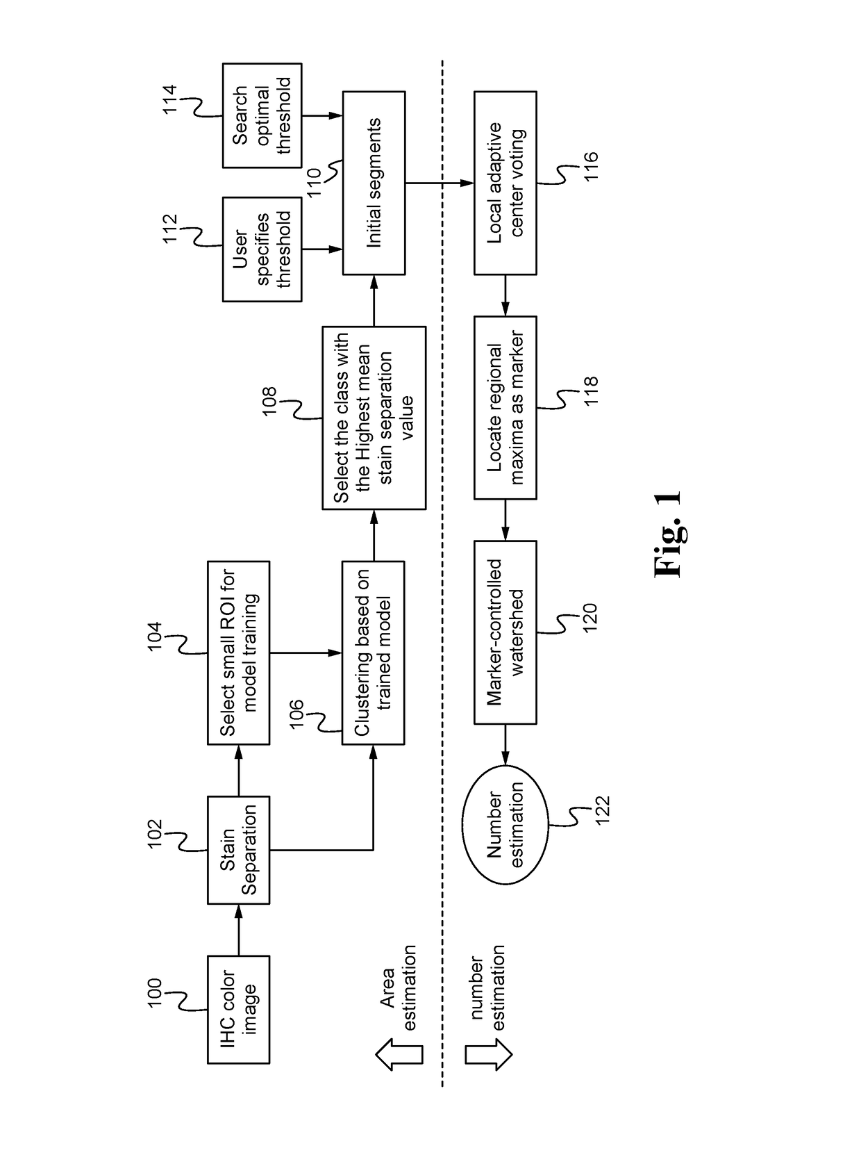
However, most of the existing research work is performing nuclei area estimation, which is fast in speed but does not provide nuclei number estimation.
Touching cases (e.g., when nuclei are connected with each other) makes the number estimation even more challenging.
Under-estimation and over-estimation are two major issues when developing automated nuclei number estimation.

Method used


Image

  • Automated nuclei area/number estimation for ihc image analysis
  • Automated nuclei area/number estimation for ihc image analysis

Examples


Embodiment Construction

[0014]An automated nuclei area and number estimation method and system enable improved Immunohistochemistry (IHC) image analysis.

[0015]The automated nuclei area and number estimation system uses a two-stage estimation framework: nuclear area estimation (e.g., number of nuclear pixels) followed by nuclear number estimation. Nuclear area is estimated from a binarized patch or patches, and these segmented patches provide local shape features which are able to facilitate number estimation.

[0016]To better distinguish a nuclear target and artifacts, the image quality of stain separation is enhanced by performing adaptive clustering based on a user-selected Region of Interest (ROI) via model training / selection.

[0017]To estimate nuclei area, stain separation is applied to separate two dominating color components, one corresponding to positive stains and the other one corresponding to negative stains. Area estimation is performed based on each color channel by adaptive thresholding. The syst...


PUM


Abstract

Automated nuclei area / number estimation utilizes a two-stage estimation framework-area estimation first followed by number estimation. After determining area information, each local patch's shape features are able to be extracted to define a local voting rule. The resulting voting score determines the strength of each local voting peak. The number of voting peaks is exactly the number of nuclei.

Description

FIELD OF THE INVENTION[0001]The present invention relates to the field of imaging. More specifically, the present invention relates to medical imaging.BACKGROUND OF THE INVENTION[0002]Immunohistochemistry (IHC) refers to the process of detecting proteins in cells of a tissue section. IHC staining is widely used in the diagnosis of abnormal cells such as those found in cancerous tumors. Common practice in pathology laboratories is to score IHC-stained images. By indicating a tumor is negative or positive, the percentage of positively stained tumor cell nuclei is able to be reported, which is able to assist pathologists for the final scoring purpose.[0003]Some research has been done for the percentage estimation of positively stained tumor cell nuclei. The goal is achieved by using a color de-convolution algorithm for separating the staining components (diaminobenzidine and hematoxylin) and adaptive thresholding for nuclear area segmentation. The quantitative results are calibrated us...

Claims


Application Information

Patent Timeline
IPC IPC(8): G06T7/00G06K9/00G06K9/46G01N33/574
CPCG06T7/0012G06K9/00127G06T2207/30024G01N33/57496G06K9/4604G06T2207/10024G06T2207/10056G06T2207/20081G06T2207/20104G06T7/11G06T7/136G06V20/69G01N15/0205G06V20/695G06V20/698
Inventor WANG, SUXU, XUNNAKAMURA, AKIRA
Owner SONY CORP
Learn More