Automatic nuclei segmentation in histopathology images

a nucleus and image technology, applied in image analysis, image enhancement, instruments, etc., can solve the problems of time-consuming and often infeasible large-scale studies

Inactive Publication Date: 2019-02-07
OREGON HEALTH & SCI UNIV
View PDF2 Cites 37 Cited by
  • Summary
  • Abstract
  • Description
  • Claims
  • Application Information

AI Technical Summary

Benefits of technology

The patent describes a method of extracting features from images using wavelets and then clustering them using a technique called k-means. This method is more robust and effective than existing methods, resulting in more accurate image analysis.

Problems solved by technology

This is time-consuming and often infeasible for large-scale studies.

Method used


Image

  • Automatic  nuclei segmentation in histopathology images
  • Automatic  nuclei segmentation in histopathology images
  • Automatic  nuclei segmentation in histopathology images

Examples


embodiment 1

2. The system of embodiment 1, wherein the image-capture device and / or the control unit is configured to carry out one or more operations automatically.

3. The system of embodiment 1, wherein the image of the cell population is a histopathology image.

embodiment 3

4. The system of embodiment 3, wherein the histopathology image is (a) an image of hemolysin and eosin (H&E) stained tissue section, or (b) an immunohistochemical (IHC) image including labeling of a biomarker in a tissue section. Optionally, the IHC image in some embodiments is one of a series of images from a single tissue section, each image reflecting the labeling of at least one different target within the tissue.

5. The system of embodiment 1, wherein the image-capture device further is configured to: (A) determine a response of a Gabor filter based at least in part on a first input-pixel value of a first pixel of the pixels of the image; and at least one of the per-pixel feature values associated with the first pixel is the response of the Gabor filter; and / or (B) determine a response of a Haralick filter based at least in part on a first input-pixel value of a first pixel of the pixels of the image; and at least one of the per-pixel feature values associated with the first pix...

embodiment 8

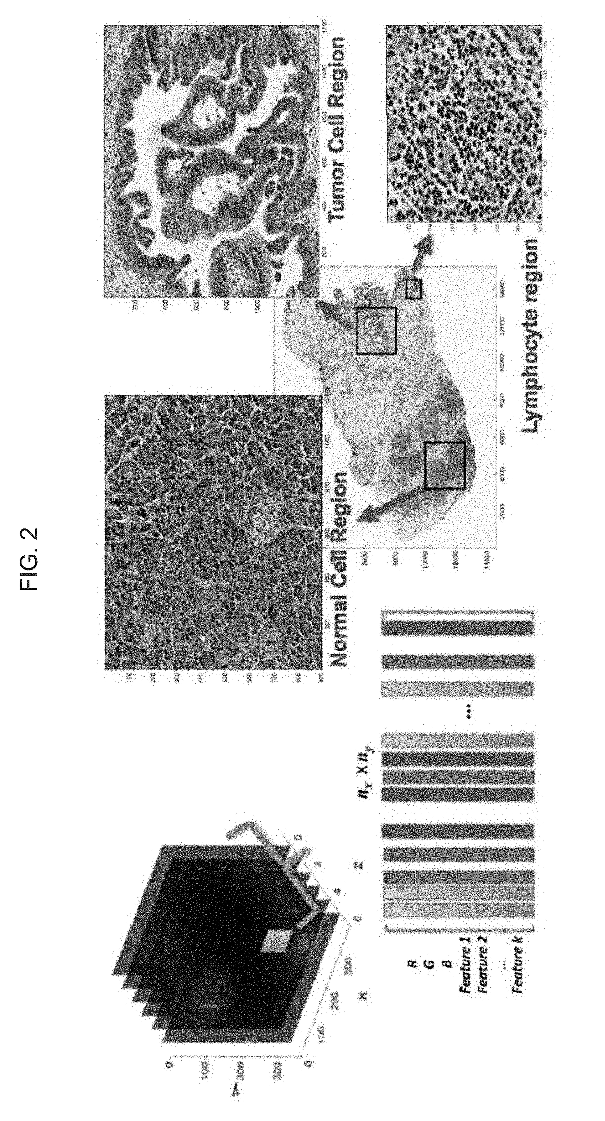
9. The system of embodiment 8, wherein at least one super-pixel includes at least one of: an R, G, B, Panchromatic (broadband), C, M, Y, Cb, Cr, CIE L*, CIE a*, CIE b*, or other data value of or determined based on a corresponding pixel; a Gabor filter response associated with a corresponding pixel; a Haralick feature value associated with a corresponding pixel; or another feature value associated with a corresponding pixel.

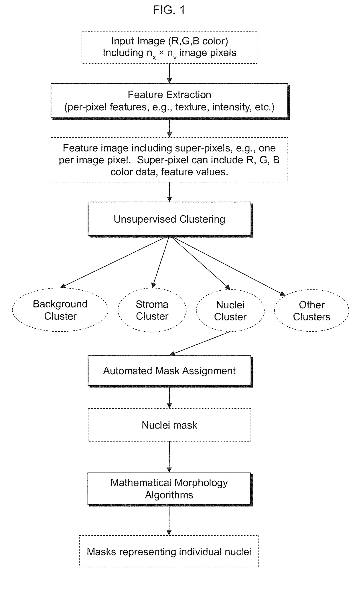
10. A computer-implemented method, including: capturing an image of a cell population, the image including input-pixel values of respective pixels of the image; determining a feature image based at least in part on the input-pixel values, the feature image including super-pixels associated with respective pixels of the pixels of the image, wherein each super-pixel includes one or more per-pixel feature value(s) associated with the respective pixel of the pixels of the image; determining a plurality of clusters based at least in part on the feature image, wherein ...


PUM


Abstract

Provided herein are systems and computer-implemented methods for quantitative analyses of tissue sections (including, histopathology samples, such as immunohistochemically labeled or H&E stained tissue sections), involving automatic unsupervised segmentation of image(s) of the tissue section(s), measurement of multiple features for individual nuclei within the image(s), clustering of nuclei based on extracted features, and / or analysis of the spatial arrangement and organization of features in the image based on spatial statistics. Also provided are computer-readable media containing instructions to perform operations to carry out such methods. A quantitative image analysis pipeline for tumor purity estimation is also described

Description

CROSS-REFERENCE TO RELATED APPLICATION[0001]This application claims the benefit of the earlier filing date of U.S. Provisional Application No. 62 / 541,475, filed Aug. 4, 2017, which earlier application is herein incorporated by reference in its entirety.FIELD[0002]Generally, this disclosure relates to image analysis, particularly analysis of cytological samples, including histochemistry such as multiplexed histochemistry. More specifically, the disclosure relates to the fields of automated cell analysis and classification.BACKGROUND OF THE DISCLOSURE[0003]In the task of grading or diagnosis of diseases in histopathology images, e.g., cancer, the identification of certain histological structures such as nuclei, lymphocytes, and glands is essential. For example, cell counts may have diagnostic significance for some cancerous conditions (Gurcan et al., Biomedical Engineering, IEEE Reviews, 2: 147-171, 2009; Irshad et al., Biomedical Engineering, IEEE Reviews, 7: 97-114, 2014). A low Gle...

Claims


Application Information

Patent Timeline
Patent Type & Authority Applications(United States)
IPC IPC(8): G06K9/00G06T7/00G06T7/11G06K9/46G06T7/44
CPCG06K9/0014G06T7/0012G06T7/11G06K9/4638G06T7/44G06T2207/10056G06T2207/30024G06T2207/10024G06T7/155G06V20/695G06V10/443G06V10/763G06F18/23213
Inventor CHANG, YOUNG HWANTHIBAULT, GUILLAUME
Owner OREGON HEALTH & SCI UNIV
Why Eureka
Try Eureka