
Method and system for radiology reporting

a radiology and report technology, applied in informatics, instruments, healthcare informatics, etc., can solve the problems of inability to standardise, inability to accept systems proposed, and high variability in report content, structure and quality

Inactive Publication Date: 2019-05-16
GRAIN IP
View PDF9 Cites 145 Cited by
  • Summary
  • Abstract
  • Description
  • Claims
  • Application Information

AI Technical Summary

Benefits of technology

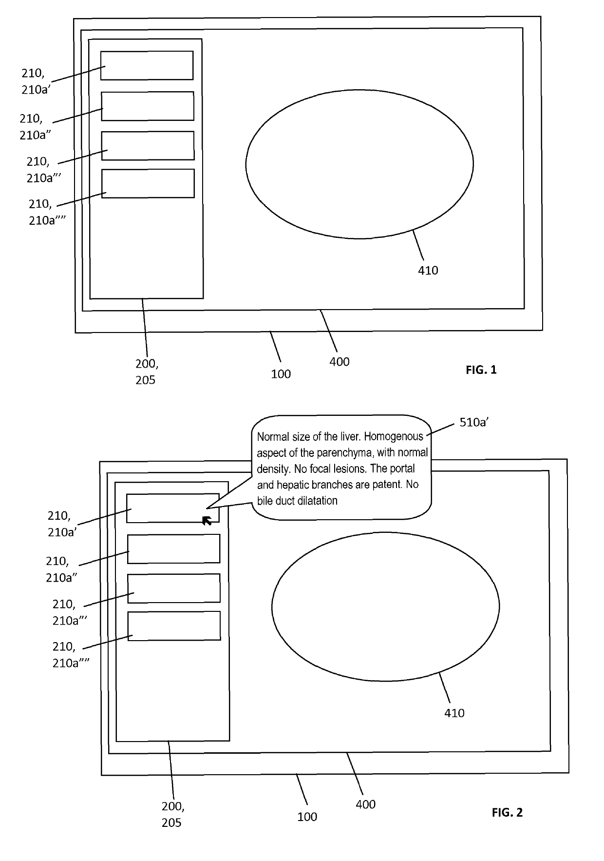
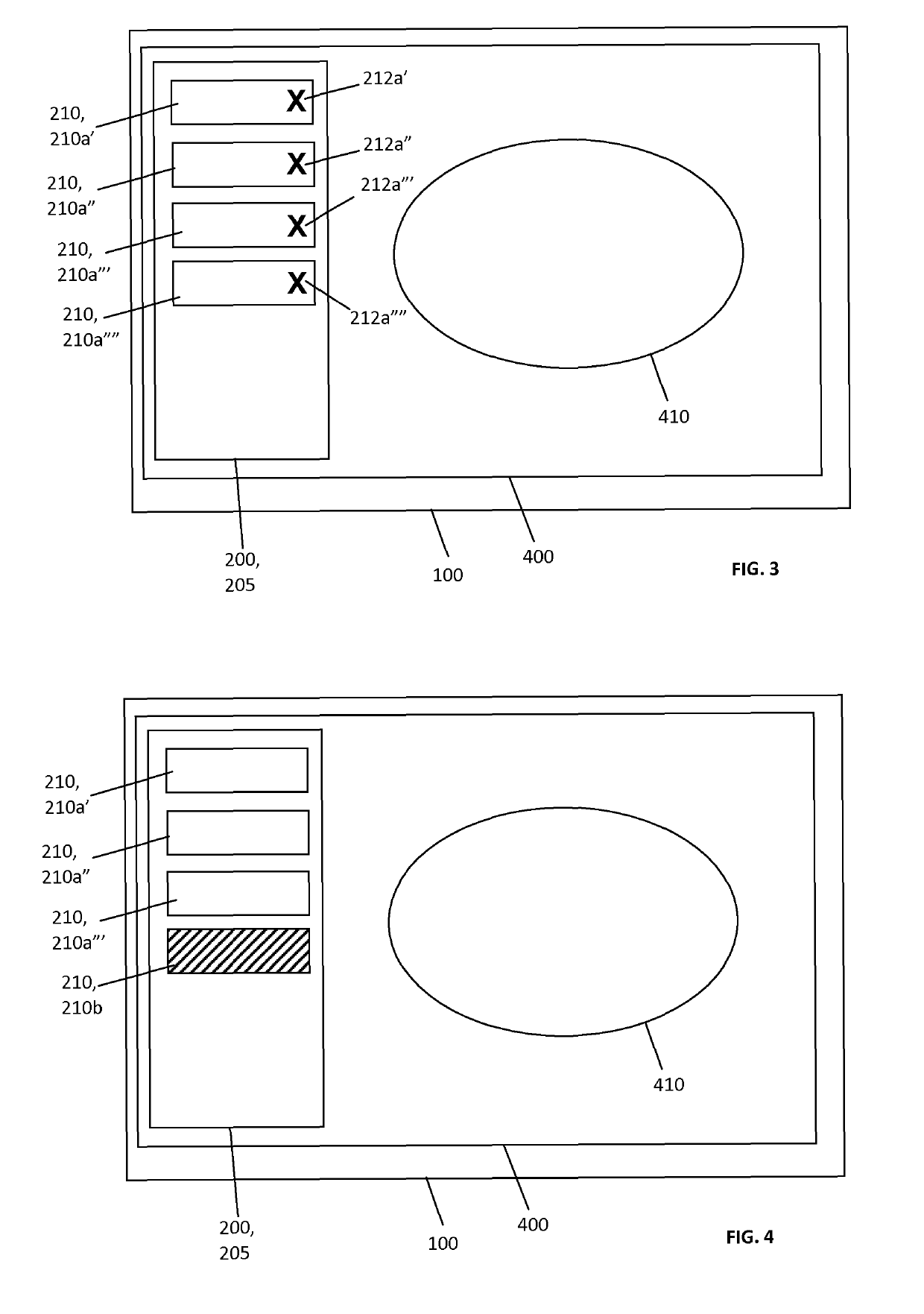
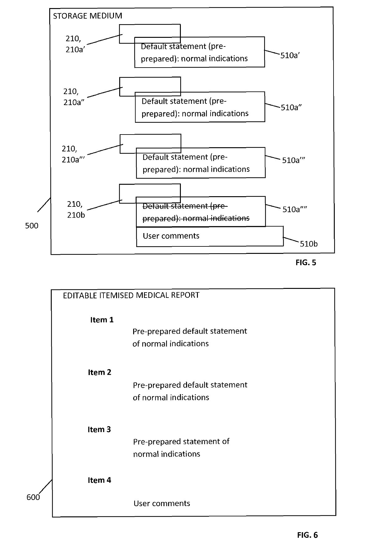
The present invention is a method for assisting a user in generating a medical report from a medical image. The method includes displaying the image and a checklist specific to the image on the screen. The checklist contains selectable items, each with a default statement indicating its normality. The user can dictate comments on the image based on the checklist items. The process generates an accurate and editable medical report with each item as a heading. This method helps the user keep track of the information and reduces oculomotor distress and improves concentration.

Problems solved by technology

Disadvantages of free dictation are well recognised and include a lack of standardisation and a high variability in report content, structure and quality.
While most experts acknowledge that making structured / standardized reports would be beneficial for quality of care, acceptance of systems proposed has been slow or even non-existent.
The reason is that SR systems tend to distract radiologists from their main task: looking at images and simultaneously dictating a report.
Switching attention between multiple-screens can induce oculomotor distress, such as headaches, eyestrain, blurred vision and general discomfort over time.

Method used


Image

  • Method and system for radiology reporting
  • Method and system for radiology reporting
  • Method and system for radiology reporting

Examples


Embodiment Construction

[0070]Before the present system and method of the invention are described, it is to be understood that this invention is not limited to particular systems and methods or combinations described, since such systems and methods and combinations may, of course, vary. It is also to be understood that the terminology used herein is not intended to be limiting, since the scope of the present invention will be limited only by the appended claims.

[0071]As used herein, the singular forms “a”, “an”, and “the” include both singular and plural referents unless the context clearly dictates otherwise.

[0072]The terms “comprising”, “comprises” and “comprised of” as used herein are synonymous with “including”, “includes” or “containing”, “contains”, and are inclusive or open-ended and do not exclude additional, non-recited members, elements or method steps. It will be appreciated that the terms “comprising”, “comprises” and “comprised of” as used herein comprise the terms “consisting of”, “consists” ...


PUM


Abstract

The present invention relates to method for assisting a user in generating an itemised medical report from at least one medical image. The at least one medical image is displayed in a display area of single computer display unit. In the same display area, a sub-region containing a checklist specific to the one or more images is displayed. Items from the checklist can be de-selected. This checklist comprises a list of selectable items, each item representing an organ, structure, or abnormality that has to be checked by the user. Linked to each item in the checklist is a default statement that is a pre-prepared statement indicative of the normality of the item. The default statement is not displayed by default as part of the checklist. The user can select an item from the checklist for providing comments by dictation thereon responsive to an observation in the radiological image. At the end of the image analysis, an editable itemised medical report is generated containing each item of the checklist as a heading and either the dictated comment or the default statement associated with the item as an observation. The editable itemised medical report is rapidly and accurately generated, information is confined to a single screen reducing the look-away time and general discomfort over time for the user.

Description

FIELD OF THE INVENTION[0001]The invention generally relates to generating a report of the analysis of a medical image, and in particular generating a report for radiological image files.BACKGROUND TO THE INVENTION[0002]Medical imaging techniques are commonly used for diagnosis and / or follow-up in clinical medicine. Examples of medical imaging techniques include X-Ray, CT (computed tomography), MRI (magnetic resonance imaging), mammography, ultrasonography, etc.[0003]Usually, a medical professional (typically a radiologist) analyses the images and dictates a medical imaging report. Methods and systems for generating a report are known in the art, for instance, WO 2012 / 037049 describes a teleradiology system having a report generation system that is template driven and provides access to macros specific to a study as well as previously dictated entries for particular fields to allow reuse of standard text in a contextual manner. WO 2012 / 071571 discloses a method for creating a radiolo...

Claims


Application Information

Patent Timeline
IPC IPC(8): G16H30/20G16H15/00G16H10/60
CPCG16H30/20G16H15/00G16H10/60G16H30/40
Inventor VAN HOE, LIEVEN
Owner GRAIN IP
Learn More