

Method and apparatus for cardiac radiological examination in coronary angiography
a radiological examination and coronary artery technology, applied in the field of medical imaging in cardiology, can solve the problems of long and laborious protocol as a whole, short time allotted for coronary artery injections, and limited total quantity of contrast fluid injected, so as to achieve the effect of better use of contrast medium
- Summary
- Abstract
- Description
- Claims
- Application Information
AI Technical Summary
Problems solved by technology
Method used
Image

Examples
Embodiment Construction
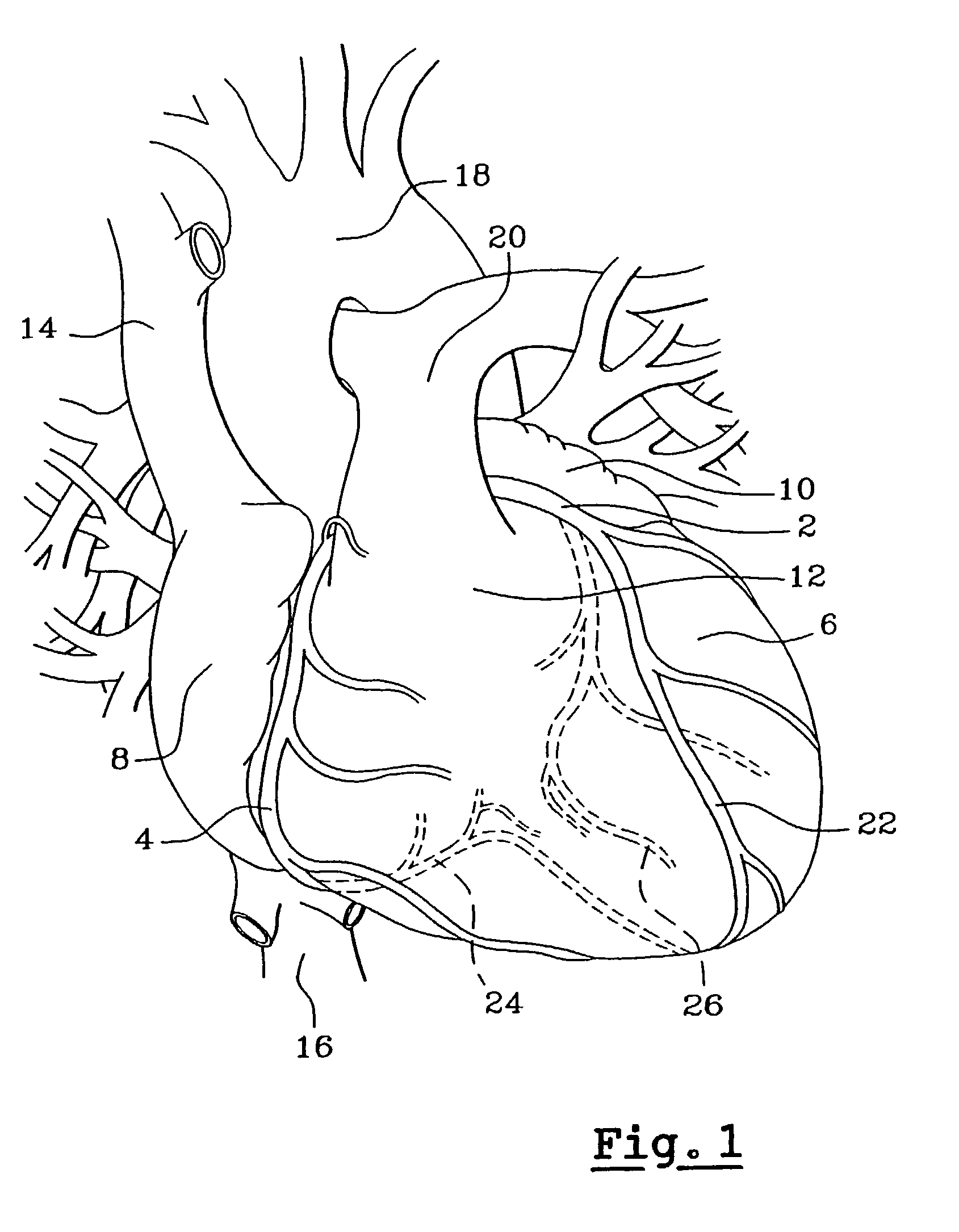
[0024]The three different catheters are positioned respectively as shown in FIG. 1: the aperture of the left coronary artery 2, the aperture of the right coronary artery 4 and the interior of the left ventricle 6.
[0025]FIG. 1 further identifies in the heart: the right auricle 8, the left auricle 10, the right ventricle 12, the superior caval vein 14, the inferior caval vein 16, the aorta 18, the pulmonary artery 20, the anterior interventricular artery 22, the posterior interventricular artery 24 and the circumflex left artery 26.
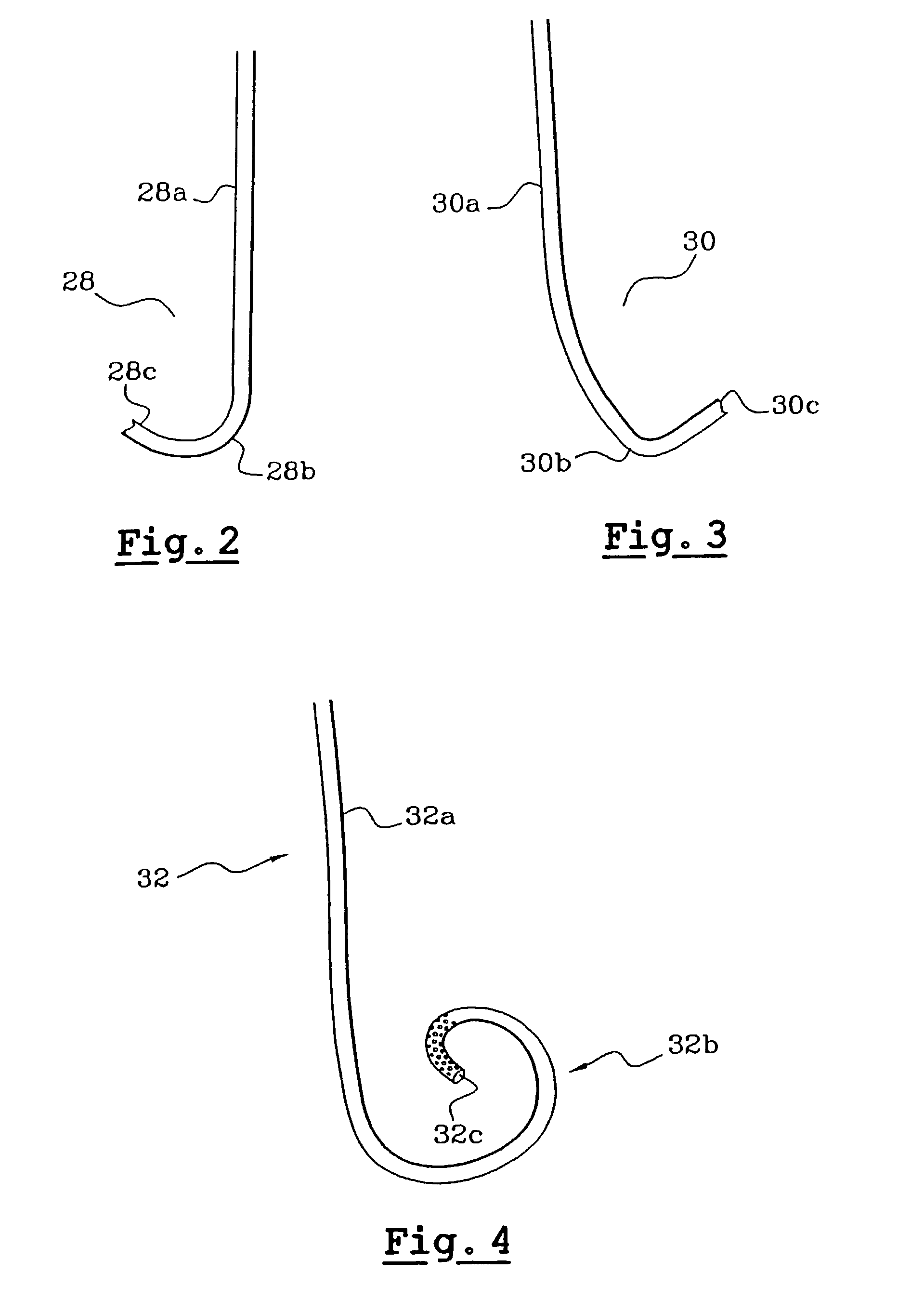
[0026]FIGS. 2, 3 and 4 respectively show the shapes that the injection ends of the three aforementioned catheters take when deployed. The catheter 28 intended for the right coronary artery as shown in FIG. 2, presents an appreciably straight section 28a which ends in an elbowed portion 28b in order to guide the tip 28c to approximately 90° from the straight part 28a, so that it can partially enter the coronary artery. The tip 28c affords a single outlet for...
PUM
 Abstract
Description
Claims
Application Information