Method and apparatus for simulating blood flow under patient-specific boundary conditions derived from an estimated cardiac ejection output

A technology of cardiac ejection and boundary conditions, applied in blood flow measurement devices, blood flow measurement, and instruments for radiological diagnosis, etc., can solve problems such as unclear pressure gradients

Inactive Publication Date: 2015-08-26
KONINKLJIJKE PHILIPS NV
View PDF4 Cites 7 Cited by
  • Summary
  • Abstract
  • Description
  • Claims
  • Application Information

AI Technical Summary

Problems solved by technology

The effect on three-dimensional (3D) blood flow may therefore not be fully assessed by such 2D measurements
The relationship between such measured areas and the physiological effects of stenotic valves (such as increased pressure gradients) is therefore unclear

Method used


Image

  • Method and apparatus for simulating blood flow under patient-specific boundary conditions derived from an estimated cardiac ejection output
  • Method and apparatus for simulating blood flow under patient-specific boundary conditions derived from an estimated cardiac ejection output
  • Method and apparatus for simulating blood flow under patient-specific boundary conditions derived from an estimated cardiac ejection output

Examples


Embodiment Construction

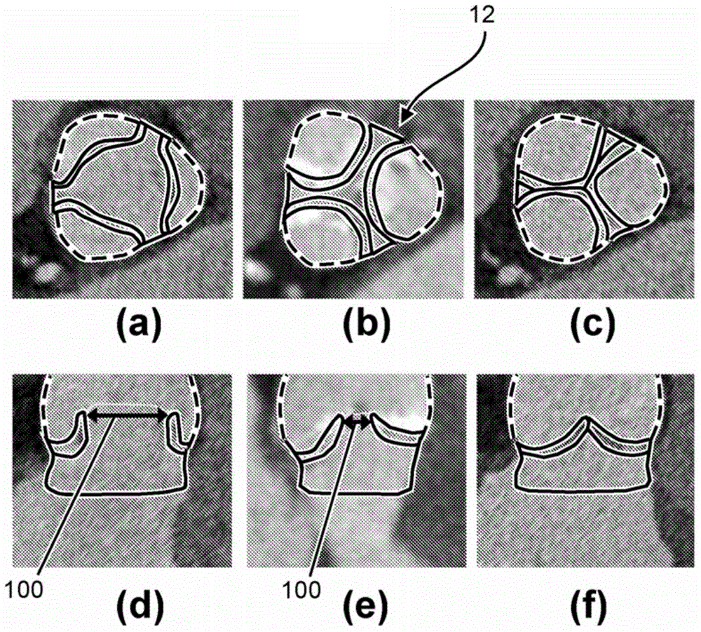
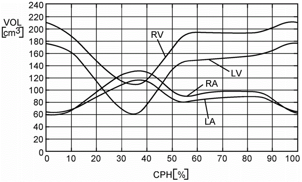
[0026] Now based on the addition of the ascending aorta through the left ventricular (LV) outflow tract (as an example of a blood cavity or cardiovascular structure close to the heart) under patient-specific boundary conditions derived from cardiac ejection output per heartbeat Embodiments are described for the simulation of blood flow in patient-specific geometries that can be derived from (at least) two images of the LV at states of maximum and minimum filling (e.g., end-diastole, end-systole) calculate. The geometry of the LV outflow tract, including the aortic root of the AV, plus ascending aorta and ventricular volumes can be obtained automatically by model-based segmentation.

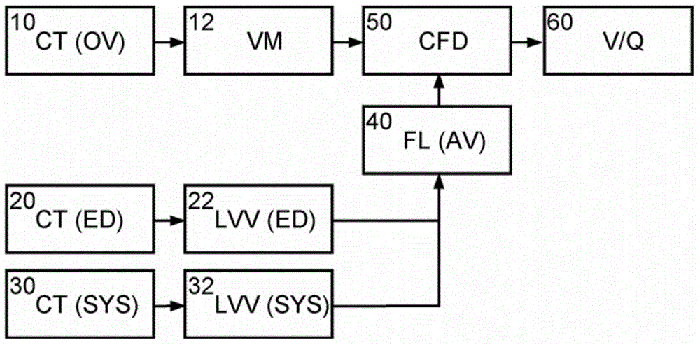
[0027] figure 1 A schematic block diagram illustrating the generation and use of patient-specific boundary conditions for simulating flow through the aortic valve is shown. figure 1 The blocks can be regarded as hardware circuits adapted to perform the corresponding functions or as steps of the ...


PUM


Abstract

The present invention relates to a method and apparatus for simulating blood flow through a cardiovascular structure, e.g. a blood cavity such as the left ventricle outflow tract, the aortic root including the AV, plus ascending aorta, a ventricle volume, the aorta or any other cavity where blood flows through, under patient-specific boundary conditions derived from the cardiac ejection output per heart stroke. The cardiac ejection output can be estimated from volumes of a heart chamber of the patient in different filling states at two or more different points in time. The results of the flow simulation can be used to derive at least one physiological parameter or can be visualized and virtual Doppler ultrasound images may be generated to allow a physician assessing the result.

Description

technical field [0001] The present invention relates to modeling the patient-specific geometry by targeting cardiovascular structures such as, but not limited to, the left ventricular outflow tract, the aortic root including the aortic valve (AV), and ascending aorta) the area of ​​blood flow. Background technique [0002] As described in "Multislice Computed Tomography for Detection of Patients With Aortic Valve Stenosis and Quantification of Severity" by G.M.Feuchtner, W.Dichtl et al., Journal of the American College of Cardiology 2006, 47(7), pp. 1410–1417, at Degenerative aortic stenosis (AS) is the second most common cardiovascular disease with an incidence of 2-7% in Western European and North American populations aged over 65 years. [0003] Management of patients with degenerative AS depends on the severity of the disease. Assessment of the severity of stenosis of the aortic valve (AV) can involve different imaging modalities. Current assessments of severity are m...

Claims


Application Information

Patent Timeline
Patent Type & Authority Applications(China)
IPC IPC(8): G06F19/00G06T19/00A61B6/00A61B8/00A61B5/026A61B5/029A61B5/055G16B5/00
CPCA61B6/5217A61B5/029A61B8/5223G06T19/00A61B6/503A61B5/0263A61B8/065G06F19/3437A61B5/055G06T2210/41G16H50/50G16B5/00A61B6/032A61B8/06A61B5/7285
Inventor J·彼得斯J·威斯H·施米特
Owner KONINKLJIJKE PHILIPS NV
Why Eureka
Try Eureka