
System, method, and computer-accessible medium for virtual pancreatography

a virtual pancreatography and computer-accessible technology, applied in the field of medical imaging, can solve problems such as difficulty in correctly identifying the type of cystic lesion by manual examination of the radiological image and no cad procedures for classifying the type of pancreatic cystic lesion

Active Publication Date: 2020-07-16
THE RES FOUND OF STATE UNIV OF NEW YORK
View PDF0 Cites 20 Cited by
  • Summary
  • Abstract
  • Description
  • Claims
  • Application Information

AI Technical Summary

Benefits of technology

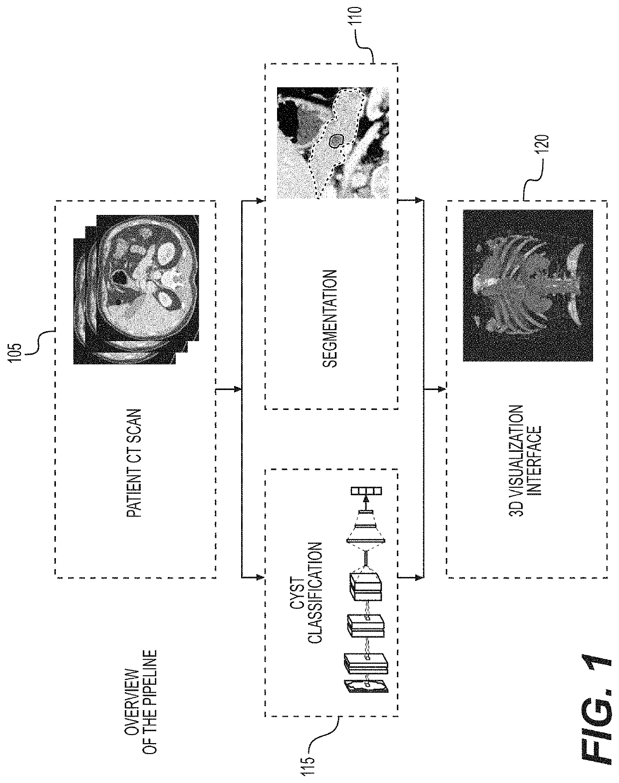
The present patent describes a system, method, and computer-accessible medium for using medical imaging data to screen for cysts. The system includes a computer-accessible medium that receives first imaging information about an organ of a patient and generates second imaging information based on the first imaging information. The second imaging information is used to segment the organ and identify different types of cysts. The system can also use a convolutional neural network to classify the cysts based on their characteristics. The system can be used to screen for cysts in a pancreas gland or other abdominal organs. Overall, the system can improve the accuracy and efficiency of identifying cysts and providing a better understanding of their characteristics.

Problems solved by technology

However, the issue can be complicated because there are many other types of pancreatic cystic lesions.
However, correctly identifying cystic lesion type by manual examination of the radiological images of pancreatic cystic lesions can be challenging, even for an experienced radiologist.
Although many procedures have been proposed for the non-invasive analysis of benign and malignant masses in various organs, there are no CAD procedures for classifying pancreatic cystic lesion type.

Method used


Image

  • System, method, and computer-accessible medium for virtual pancreatography
  • System, method, and computer-accessible medium for virtual pancreatography
  • System, method, and computer-accessible medium for virtual pancreatography

Examples


Embodiment Construction

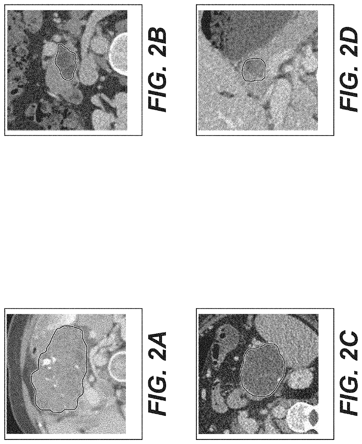
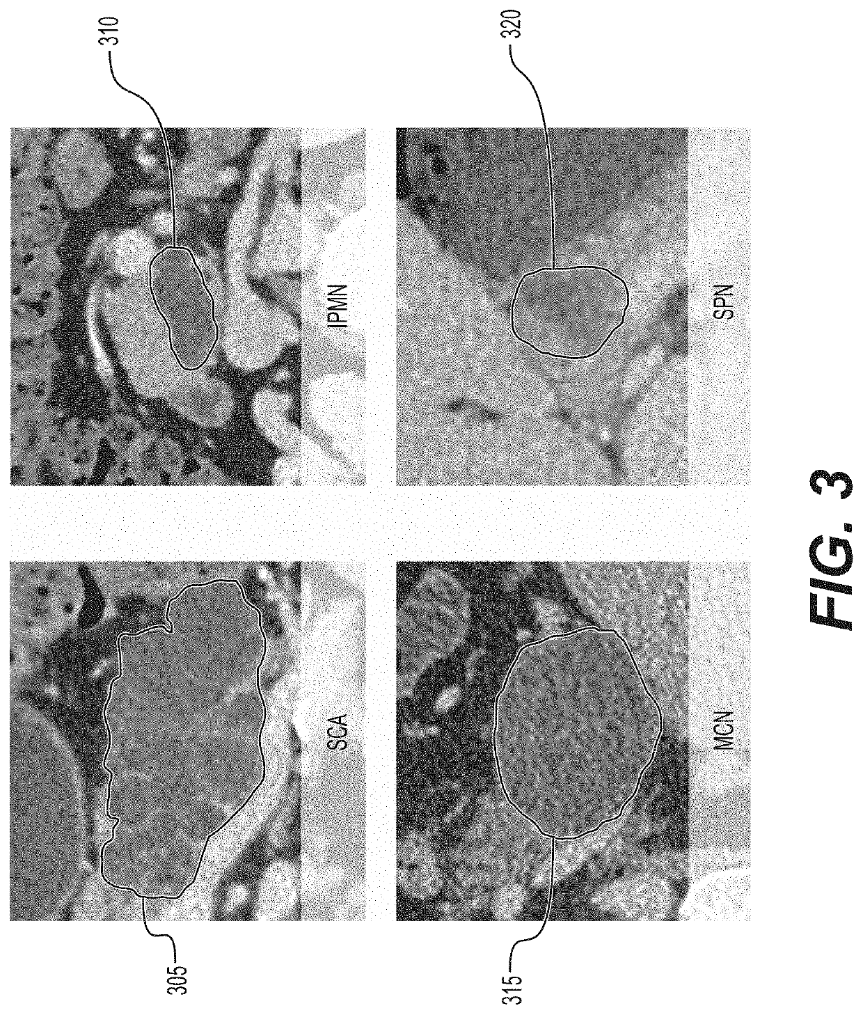
[0009]A system, method, and computer-accessible medium for using medical imaging data to screen for a cyst(s) (e.g., for segmentation and visualization of a cystic lesion) can include, for example, receiving first imaging information for an organ(s) of a one patient(s), generating second imaging information by performing a segmentation operation on the first imaging information to identify a plurality of tissue types, including a tissue type(s) indicative of the cyst(s), identifying the cyst(s) in the second imaging information, and applying a first classifier and a second classifier to the cyst(s) to classify the cyst(s) into one or more of a plurality of cystic lesion types. The first classifier can be a Random Forest classifier and the second classifier can be a convolutional neural network classifier. The convolutional neural network can include at least 6 convolutional layers, where the at least 6 convolutional layers can include a max-pooling layer(s), a dropout layer(s), and ...


PUM


Abstract

A system, method, and computer-accessible medium for using medical imaging data to screen for a cystic lesion(s) can include, for example, receiving first imaging information for an organ(s) of a one patient(s), generating second imaging information by performing a segmentation operation on the first imaging information to identify a plurality of tissue types, including a tissue type(s) indicative of the cystic lesion(s), identifying the cystic lesion(s) in the second imaging information, and applying a first classifier and a second classifier to the cystic lesion(s) to classify the cystic lesion(s) into one or more of a plurality of cystic lesion types. The first classifier can be a Random Forest classifier and the second classifier can be a convolutional neural network classifier. The convolutional neural network can include at least 6 convolutional layers, where the at least 6 convolutional layers can include a max-pooling layer(s), a dropout layer(s), and fully-connected layer(s).

Description

CROSS-REFERENCE TO RELATED APPLICATION(S)[0001]This application relates to and claims priority from U.S. Patent Application No. 62 / 524,819, filed on Jun. 26, 2017, the entire disclosure of which is incorporated herein by reference.STATEMENT REGARDING FEDERALLY SPONSORED RESEARCH[0002]This invention was made with government support under Grant Nos. CNS-0959979, IIP 1069147, and CNS-1302246, awarded by the National Science Foundation. The government has certain rights in the invention.FIELD OF THE DISCLOSURE[0003]The present disclosure relates generally to medical imaging, and more specifically, to exemplary embodiments of an exemplary system, method, and computer-accessible medium for virtual pancreatography.BACKGROUND INFORMATION[0004]Pancreatic cancer, or pancreatic ductal adenocarcinoma (“PDAC”) as it can be formally known, can be one of the most lethal of all cancers with an extremely poor prognosis and an overall five-year survival rate of less than 9%. There are no specific ear...

Claims


Application Information

Patent Timeline
Patent Type & Authority Applications(United States)
IPC IPC(8): G06T7/00G06T7/11G06N20/20G06N3/04G06V10/764
CPCG06T7/0012G06N20/20G06T2207/30096G06T7/11G06N3/0454G06T2207/20081G06T7/194G06T7/187G06T2207/10081G06T2207/10088G06T2207/20076G06T2207/20104G06T2207/20101G06T2207/30056G06T2207/20128G06T2207/20084G06N3/082G06V10/454G06V2201/10G06V2201/03G06V10/764G06V10/809G06N5/01G06N7/01G06N3/048G06N3/045G06F18/2413G06F18/24323G06F18/254
Inventor KAUFMAN, ARIEDMITRIEV, KONSTANTIN
Owner THE RES FOUND OF STATE UNIV OF NEW YORK
Learn More