Device for training users of an ultrasound imaging device

a technology of ultrasound imaging and training users, applied in the field of medical simulators, can solve the problems of simulators often insufficient, inexperienced doctors and technicians are typically not trained to use obstetric sonography to detect fetal abnormalities, and inability to identify such abnormalities

Inactive Publication Date: 2020-12-24
ARIEL UNIV RES & DEV
View PDF0 Cites 0 Cited by
  • Summary
  • Abstract
  • Description
  • Claims
  • Application Information

AI Technical Summary

Benefits of technology

[0064]In some embodiments the first virtual volume simulates a uterine volume containing amniotic fluid and the second virtual volume simulates an embryo or fetus, and the user-assessment module is configured to train the user to perform an amniocentesis procedure without harming the embryo or fetus.

Problems solved by technology

Technicians and doctors are typically not trained to use obstetric sonography to detect fetal abnormalities.
Thus, inexperienced doctors and technicians are typically incapable of identifying such abnormalities when these are encountered in practice.
Such simulators are often insufficient because they fail to simulate motion of muscles during the procedure, or various types of abnormalities that can be encountered during the sonography.
Such simulators are insufficient since the fetus model is static, and such training simulators fail to simulate an important factor of obstetric sonography, fetal movement.
Further, in such training simulators, the maternal and embryo features are normal and therefore useless for training in identifying fetal abnormalities.

Method used


Image

  • Device for training users of an ultrasound imaging device
  • Device for training users of an ultrasound imaging device
  • Device for training users of an ultrasound imaging device

Examples


Embodiment Construction

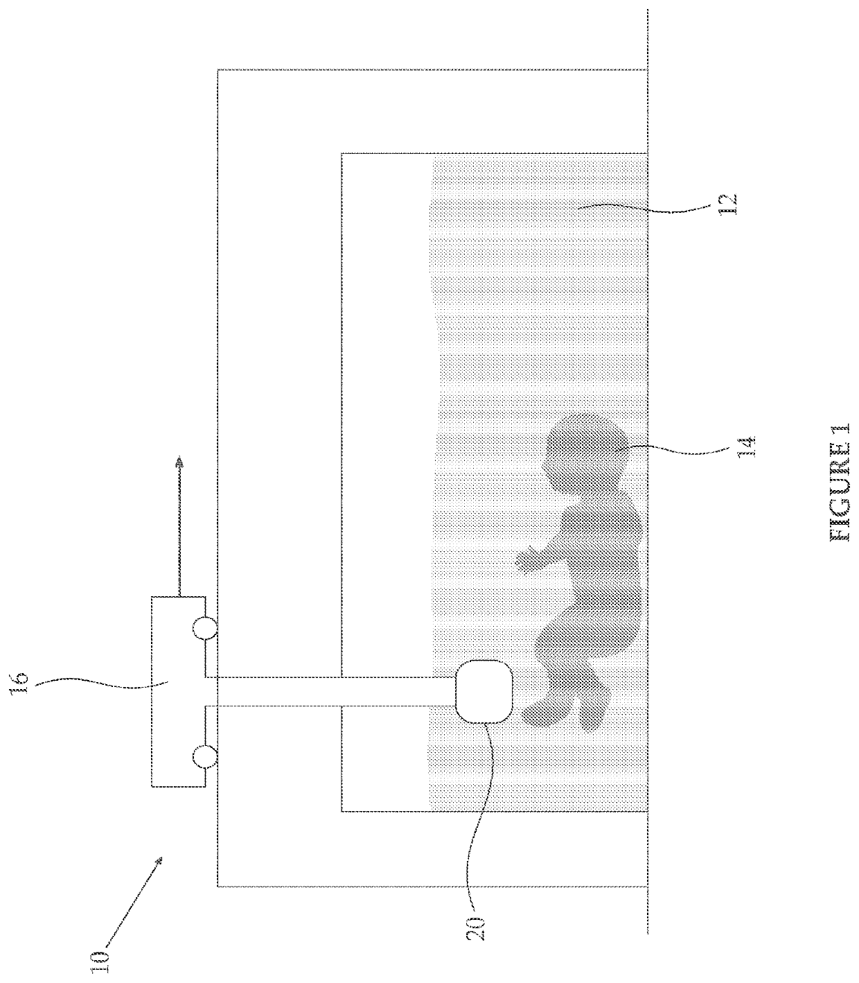
[0123]The invention, in some embodiments, relates to the field of medical simulators, and more particularly, in some embodiments, to methods and devices for training ultrasound users to perform medical sonography, such as gynaecological sonography, cardiological sonography, gasteroenterological sonography, neurological sonography, musculoskeletal sonography, and CT scans, and to identify abnormalities seen in such tests.

[0124]As discussed above, methods and devices are needed in order to train users such as doctors and ultrasound technicians to recognize abnormalities and anomalies, such as embryonic abnormalities, or to safely guide medical devices, such as amniocentesis needles, using ultrasound imaging.

[0125]The principles, uses and implementations of the teachings of the invention may be better understood with reference to the accompanying description and figures. Upon perusal of the description and figures present herein, one skilled in the art is able to implement the teaching...


PUM


Abstract

Disclosed are methods and devices for simulating ultrasound procedures and for training ultrasound users. Additionally disclosed are methods and devices for simulating needle insertion procedures, such as amniocentesis procedures, and for training physicians to perform such needle insertion procedures.

Description

RELATED APPLICATION[0001]This application is a continuation of U.S. patent application Ser. No. 14 / 387,548 filed on Sep. 24, 2014, which is a National Phase of PCT Patent Application No. PCT / IB2013 / 052581 having International Filing Date of Mar. 31, 2013, which claims the benefit of priority of U.S. Provisional Patent Application No. 61 / 618,791 filed on Apr. 1, 2012. The contents of the above applications are all incorporated by reference as if fully set forth herein in their entirety.FIELD AND BACKGROUND OF THE INVENTION[0002]The invention, in some embodiments, relates to the field of medical simulators, and more particularly, in some embodiments, to methods and devices for training ultrasound users for example, to perform medical sonography or needle-insertion procedures.[0003]Ultrasound is a cyclic pressure wave with a frequency greater than about 20000 Hz, the upper limit of human hearing.[0004]In sonography, such as medical sonography, ultrasound is used for imaging, especially...

Claims


Application Information

Patent Timeline
Patent Type & Authority Applications(United States)
IPC IPC(8): G09B23/28G06T19/00A61B10/00
CPCG09B23/286G06T2215/16G06T2219/008G06T19/00A61B10/0048
Inventor TEPPER, RONNIESHVALB, NIRBEN-MOSHE, BOAZ
Owner ARIEL UNIV RES & DEV
Why Eureka
Try Eureka