System, method, and computer-accessible medium for processing brain images and extracting neuronal structures

a brain image and neuronal structure technology, applied in the field of microscopy, can solve the problems of poor axial resolution, degraded contrast between foreground and background voxels, and difficulty in analysis and visualization of wf data, and achieve the effect of minimizing an effect, maximizing the effect of in-focus voxels, and minimizing an

Pending Publication Date: 2021-02-18
THE RES FOUND OF STATE UNIV OF NEW YORK
View PDF4 Cites 2 Cited by
  • Summary
  • Abstract
  • Description
  • Claims
  • Application Information

AI Technical Summary

Benefits of technology

[0015]In certain exemplary embodiments of the present disclosure, the gradient-based distance transform can include determining the global minimum intensity value for each z-slice in the first imaging information. The imaging information can be split into a plurality of tiles, the second imaging information can be generated by separately applying the gradient-based distance transform to each of the tiles, and the image(s) can be generated by stitching together the second imaging information. The gradient-based distance transform can be applied to identify (i) in-focus voxels in the first imaging information and (ii) out-of-focus voxels in the first imaging information, minimize an effect of the out-of-focus voxels, and maximizing the effect of the in-focus voxels. The image(s) can be gigapixel image(s) having a 360° horizontal field-of-view. The image(s) can include (i) a bounded view of the anatomical structure(s), (ii) a structural view of the anatomical structure(s), or (iii) a classification view of the anatomical structure(s). The anatomical structure(s) can be a neurite(s) or a cell body(ies). The biological sample(s) can be brain tissue.
[0016]Additionally, an exemplary system, method, and computer-accessible medium for generating an image(s) of an anatomical structure(s) in a biological sample(s) can include receiving wide-field microscopy imaging information for the biological sample, identifying (i) in-focus voxels in the imaging information and (ii) out-of-focus voxels in the imaging information, minimizing an effect of the out-of-focus voxels with a penalized voxel weighting, maximizing the effect of the in-focus voxels, and generating the image(s) based on the minimized out-of-focus voxels and the maximized in-focus voxels.

Problems solved by technology

As a result, the acquired images can suffer from degraded contrast between foreground and background voxels due to out-of-focus light swamping the in-focus information, low signal-to-noise ratio, and poor axial resolution.
Thus, analysis and visualization of WF data can be a challenge for domain experts.
Transfer function designs for the volume rendering of microscopy images (see, e.g., References 6 and 53), do not yield effective results when applied to WFM images.
However, they are complex and time-consuming to compute, often requiring rounds of iterative approximations to produce the corrected image (see, e.g., Reference 45), and depend on detailed parameter inputs.
These limitations compel neurobiologists to use rudimentary methods, such as manually traversing 2D slices of the volumetric image, or maximal intensity projections, for better visibility of features at the cost of losing 3D information.
However, due to out-of-focus light spreading through the WFM data and its poor axial resolution, the naïve application of these procedures on WFM data, may not produce effective visualizations.
This makes transfer function adjustment an arduous task for neurobiologists.
However, overlapping intensity ranges of the out-of-focus light and neuronal structures make this procedure inapplicable to WFM datasets.
The optical arrangement of a WF microscope lacks the capability to reject out-of-focus light emitted by fluorescent-tagged biological targets.
However, it can depend on a detailed set of microscopy parameters and is subject to changes in the experimental procedure.
Even though deconvolution is an effective procedure for restoring microscopy images, the time and memory requirements to process large microscopy images make them less practical for regular use by domain experts.

Method used


Image

  • System, method, and computer-accessible medium for processing brain images and extracting neuronal structures
  • System, method, and computer-accessible medium for processing brain images and extracting neuronal structures
  • System, method, and computer-accessible medium for processing brain images and extracting neuronal structures

Examples


Embodiment Construction

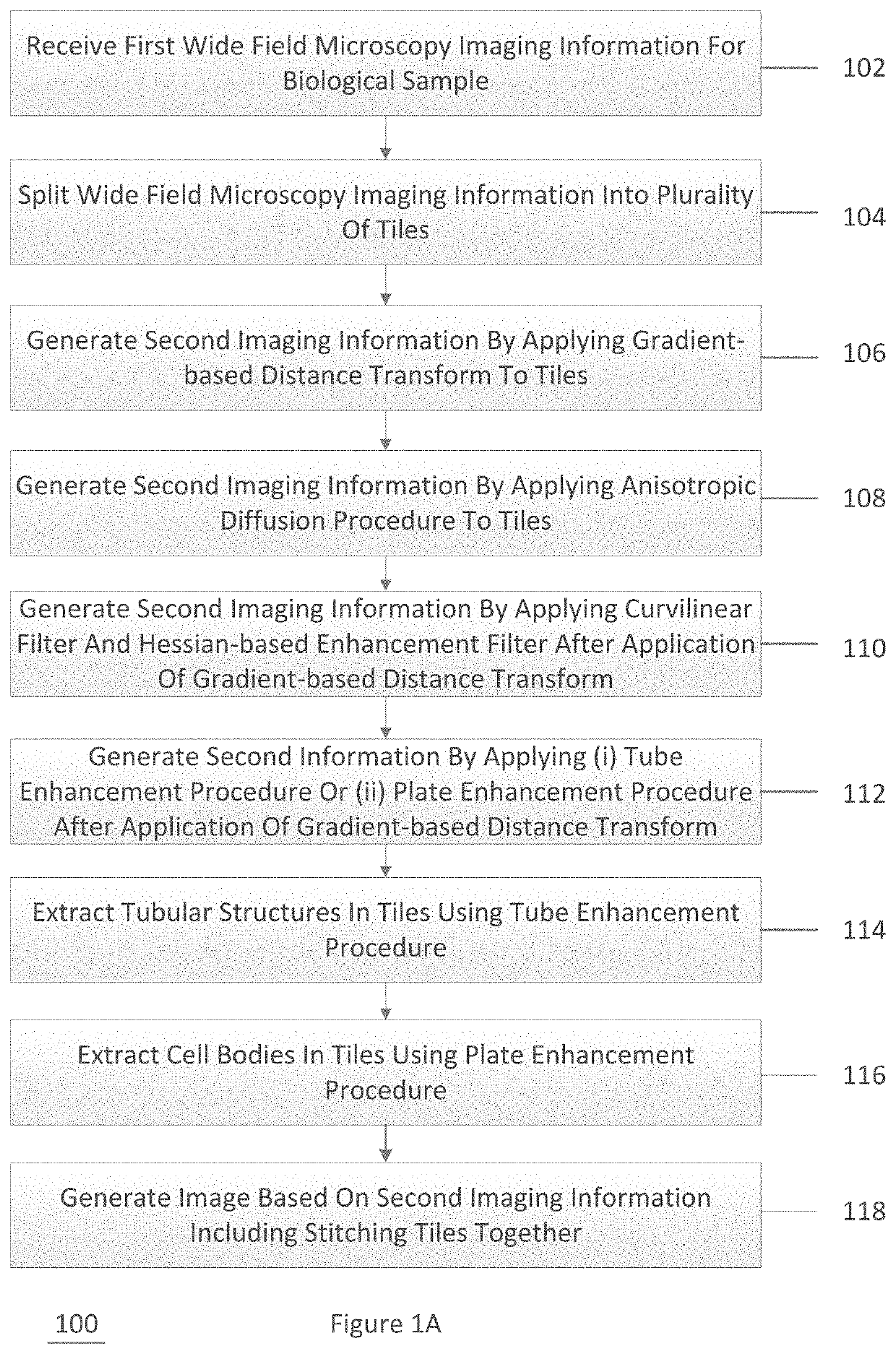
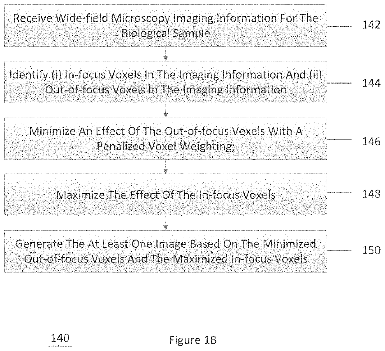
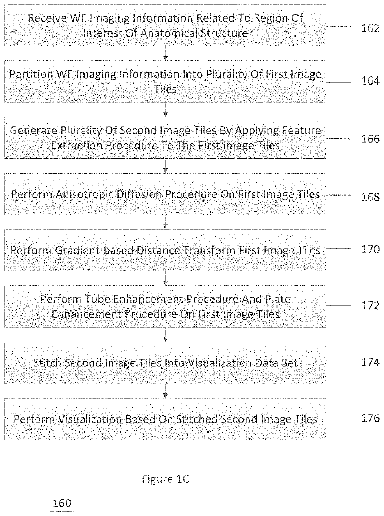
[0013]An exemplary system, method, and computer-accessible medium for generating an image(s) of an anatomical structure(s) in a biological sample(s) can include receiving first wide field microscopy imaging information for the biological sample, generating second imaging information by applying a gradient-based distance transform to the first imaging information, and generating the image(s) based on the second imaging information. The second imaging information can be generated by applying an anisotropic diffusion procedure to the first imaging information. The second imaging information can be generated by applying a curvilinear filter and a Hessian-based enhancement filter after the application of the gradient-based distance transform. The second information can be generated by applying (i) a tube enhancement procedure or (ii) a plate enhancement procedure after the application of the gradient-based distance transform. Tubular structures in the first imaging information can be ext...


PUM


Abstract

An exemplary system, method, and computer-accessible medium for generating an image(s) of an anatomical structure(s) in a biological sample(s) can include receiving first wide field microscopy imaging information for the biological sample, generating second imaging information by applying a gradient-based distance transform to the first imaging information, and generating the image(s) based on the second imaging information. The second imaging information can be generated by applying an anisotropic diffusion procedure to the first imaging information. The second imaging information can be generated by applying a curvilinear filter and a Hessian-based enhancement filter after the application of the gradient-based distance transform. The second information can be generated by applying (i) a tube enhancement procedure or (ii) a plate enhancement procedure after the application of the gradient-based distance transform.

Description

CROSS-REFERENCE TO RELATED APPLICATION(S)[0001]This application relates to and claims priority from U.S. Patent Application No. 62 / 887,719, filed on Aug. 16, 2019, the entire disclosure of which is incorporated herein by reference.STATEMENT REGARDING FEDERALLY SPONSORED RESEARCH[0002]This invention was made with government support under Grant No. CNS1650499, awarded by the National Science Foundation and Grant No. HL127522, awarded by the National Institutes of Health. The government has certain rights in the invention.FIELD OF THE DISCLOSURE[0003]The present disclosure relates generally to microscopy, and more specifically, to exemplary embodiments of an exemplary system, method, and computer-accessible medium for processing brain images and extracting neuronal structures with wide-field microscopy.BACKGROUND INFORMATION[0004]The understanding of neural connections that underline brain function is central to neurobiology research. Advances in microscopy technology have been instrum...

Claims


Application Information

Patent Timeline
Patent Type & Authority Applications(United States)
IPC IPC(8): G06K9/00G06T7/00
CPCG06K9/0014G06T7/0012G06T2207/30024G06T2207/20021G06T2207/30016G06T2207/10056G06T2207/20041G06V20/695
Inventor KAUFMAN, ARIEBOORBOOR, SAEED
Owner THE RES FOUND OF STATE UNIV OF NEW YORK
Why Eureka
Try Eureka