However, current CT technology is limited by a trade-off between high longitudinal resolution and fast volume scanning.
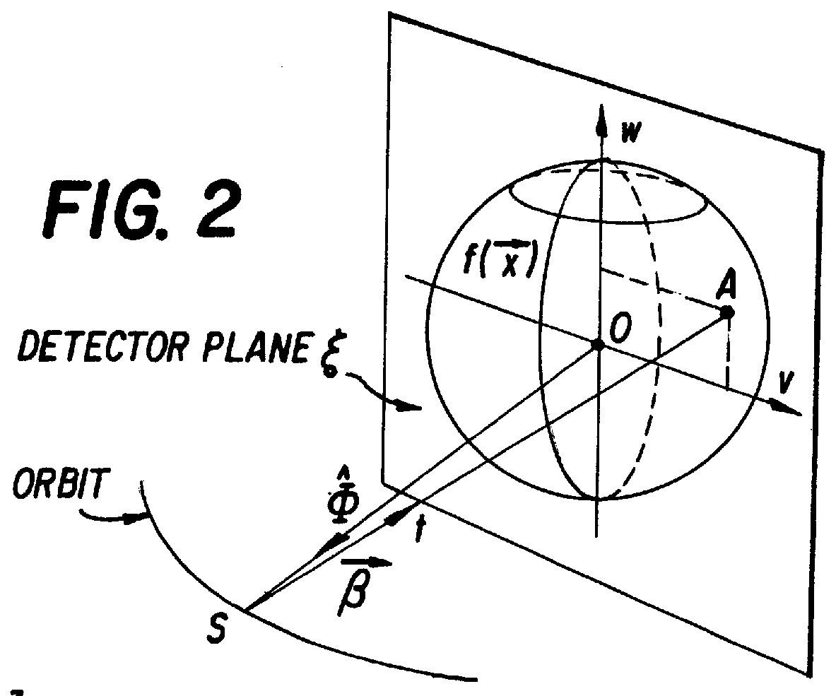
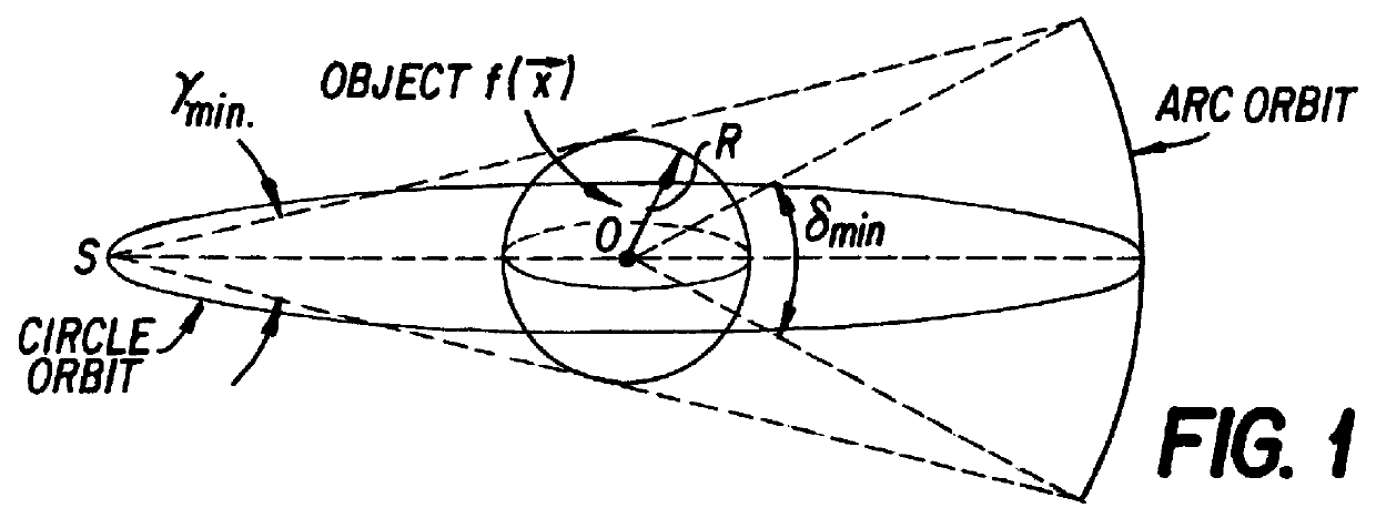
Single circle cone-beam geometry, in which the source always lies on a circle, cannot provide a complete set of data to exactly reconstruct the object.
's algorithm causes some unavoidable distortion in the non-central transverse planes, as well as resolution degradation in the longitudinal direction.
Thus, errors can be introduced by the implementation of the Gangreat method that can be greater than those produced using the Feldkamp, et al. method and such errors are not correlated with the cone-beam angle.
However, such an integer requirement condition is too restrictive for practical application since the only known source point geometry which meets that condition is a straight line.
However, that technique requires the removal of redundant and unnecessary data, which necessarily requires more computing time and complexity than the method and system of the present invention.
It cannot result in exact reconstruction and it is not acceptable in many clinical applications when the cone angle is large.
The incompleteness of the projection data results in some unavoidable blurring in the planes away from the central z plane and a resolution loss in the z direction (i.e., Feldkamp, et al.
The reconstruction error due to the incompleteness of the projection data could be up to 50% of the signal when using Feldkamp, et al.
Projection images using such standard projectional techniques do not provide sufficient information with which to detect and completely characterize all vascular lesions.
That lack of complete data impairs the ability of the physician to determine the optimal therapeutic procedure.
Obviously, an inappropriate choice of intervention based on improper knowledge of the patient's anatomy can lead to unnecessary interventions, a sub-optimal outcome, injury or death.
Second, IA-DSA images are of reduced usefulness due to vessel overlap, particularly when non-selective injections are used.
However, even with multiple views, the number of views is limited, which often results in non-detected lesions because of the failure to achieve orthogonal projection and overlap.
Consequently, the angiographic procedure can become prolonged, increasing patient morbidity from lengthened catherterization time, increasing contrast as well as the radiation dose, while also increasing procedure costs.
There is also an added risk of complications related to percutaneous cannulation of an artery and the manipulation of the IA catheters and wires in critical vessels which are often affected by vascular disease.
The risk of procedure related vascular injury and stroke is also present, with major morbidity.
Moreover, such angiogr

