Cytological evaluation of breast duct epithelial cells retrieved by ductal lavage

a cytological evaluation and breast duct technology, applied in the field of human breast duct epithelial cells evaluation by cytology, can solve the problems of insufficient cytological yield, inconvenient collection of samples, and inability to obtain sufficient cytological yield, etc., to achieve consistent, uniform, respected diagnostic tools

Inactive Publication Date: 2004-01-06
ATOSSA THERAPEUTICS INC
View PDF5 Cites 14 Cited by
  • Summary
  • Abstract
  • Description
  • Claims
  • Application Information

AI Technical Summary

Problems solved by technology

Usually several punctures with an aspiration needle are needed to locate a target lesion; tracks are created with the withdrawal of cancerous cells through the breast with a coordinate risk of spreading the cancer cells to the blood stream along the tracks; scarring can occur at the sites of entry; the punctures can be painful; multiple punctures can result in a sore breast for a time after the procedure; often multiple entries are needed to locate a lesion that provides a positive sample.
So described previous attempts at obtaining ductal fluid including nipple aspiration at the nipple surface, either with or without first infusing fluid into the duct, and early attempts at obtaining fluid oozing from the nipple surface after removing the fluid infusion catheter from the duct post fluid infusion have been found inadequate to obtain sufficient cellular yields for a cytological analysis.
These samples were not found to be a consistent, uniform, and respected diagnostic tool of breast cancer.
Likewise cell yields from these methods using a catheter for fluid infusion of about 1.5 ml of wash fluid, and where the catheter is withdrawn prior to collection of any expressed fluid on the nipple surface, have not yielded cell counts much improved from traditional nipple aspiration.

Method used


Image

  • Cytological evaluation of breast duct epithelial cells retrieved by ductal lavage
  • Cytological evaluation of breast duct epithelial cells retrieved by ductal lavage

Examples


Embodiment Construction

The following preferred embodiments and examples are offered by way of illustration and not by way of limitation.

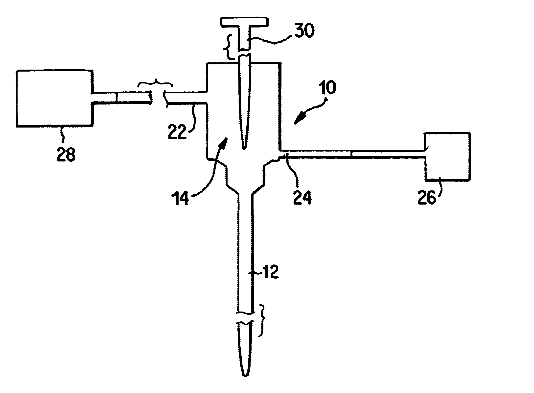
FIG. 1 illustrates an example of a tool or apparatus 10 for accessing a breast duct and collecting breast duct fluid according to an aspect of the present invention. The tool 10 comprises an elongated single lumen 12 for positioning within the breast duct and infusing and collecting fluid from within the breast duct. The tool 10 also includes a fluid infusion and collection hub 14 that is in fluid communication with the lumen 12. The hub 14 includes an infusion port 24 through which fluid from reservoir 26 is introduced into the hub 14, the lumen 12 and eventually the breast duct. The hub 14 also comprises a collection port 22 which is connected to a collection receptacle 28. Reservoir 26 and receptacle 28 can each comprise a syringe or other well known fluid carrying container. A probe 30 can be used to introduce the lumen 12 into the breast duct or locate a ductal openi...


PUM

PropertyMeasurementUnit
diameteraaaaaaaaaa
diameteraaaaaaaaaa
volumesaaaaaaaaaa

Abstract

Methods and systems for cytologically evaluating breast duct epithelial cells retrieved by ductal lavage by morphology and other parameters.

Description

1. Field of the InventionThe field of this invention is evaluating human breast duct epithelial cells by cytology to identify breast cancer and breast precancer.2. Description of the Background ArtBreast cancer is believed to originate in the lining of a single breast milk duct in the breast; and additionally human breasts are believed to contain from 6 to 9 of these ducts. See Sartorius, JAMA 224 (6): 823-827 (1973). For several decades significant members of the medical community dedicated to studying breast cancer have believed and shown that the cytological analysis of cells retrieved from nipple discharge from the breast milk ducts can provide valuable information leading to identifying patients at risk for breast cancer. Indeed, Papanicolaou contributed to the genesis of such a possibility of a "Pap" smear for breast cancer by analyzing the cells contained in nipple discharge. See Papanicolaou et al, "Exfoliative Cytology of the Human Mammary Gland and Its Value in the Diagnos...

Claims


Application Information

Patent Timeline
Patent Type & Authority Patents(United States)
IPC IPC(8): G01N33/50G01N33/574C12N15/09G01N33/48C12Q1/02C12Q1/68
CPCG01N33/5005G01N33/57415
Inventor SOITO, ANGELALJUNG, BRITT-MARIECHEW, KAREN
Owner ATOSSA THERAPEUTICS INC
Why Eureka
Try Eureka