Visceral anastomotic device and method of using same

an anastomosis and visceral technology, applied in the field of visceral anastomosis devices and methods, can solve the problems of insufficiently simplified anastomosis methods, above methods still require a great deal of effort in their use, and achieve the effect of ensuring the effect of reconnection, and ensuring the strength of the anastomosis

Inactive Publication Date: 2005-08-09
CITY OF HOPE
View PDF19 Cites 72 Cited by
  • Summary
  • Abstract
  • Description
  • Claims
  • Application Information

AI Technical Summary

Benefits of technology

[0011]The stent used in the present invention is comprised of a biocompatible material, such as polyglycolic acid (“PGA”), in the form of a semi-rigid tubular structure. Means are provided integral to the stent's construction whereby it is given flexibility without compromising its strength and therefore its ability to serve as an effective frame for anastomosis. Therefore, the surgeon is provided with a flexible stent which allows the luminal stumps to be more easily reconnected by the use of sutures, surgical staples, biocompatible sealants, or other known techniques. The advantages of such a stent are especially apparent when the anastomosis is performed by laparoscopic techniques.
[0012]A stent of the present invention, and the accompanying method of its use, can be employed in the anastomosis of various hollow organs including the intestines, bowel, bile duct, ureter, and fallopian tubes. The outside diameter and thickness of the stent can be varied such that it can provide sufficient structural strength and hold the stumps of the luminal viscus snugly in place without increasing the difficulty in manipulating the stent.

Problems solved by technology

The above mentioned attempts have been unsuccessful in providing an adequately simplified method for anastomosis.
The above methods still require a great deal of effort in their use, and their designs make them quite restrictive when used in tight quarters such as during laparoscopic procedures.

Method used


Image

  • Visceral anastomotic device and method of using same
  • Visceral anastomotic device and method of using same

Examples


Embodiment Construction

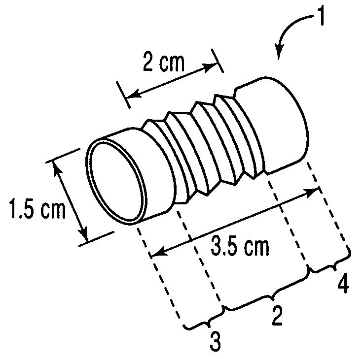
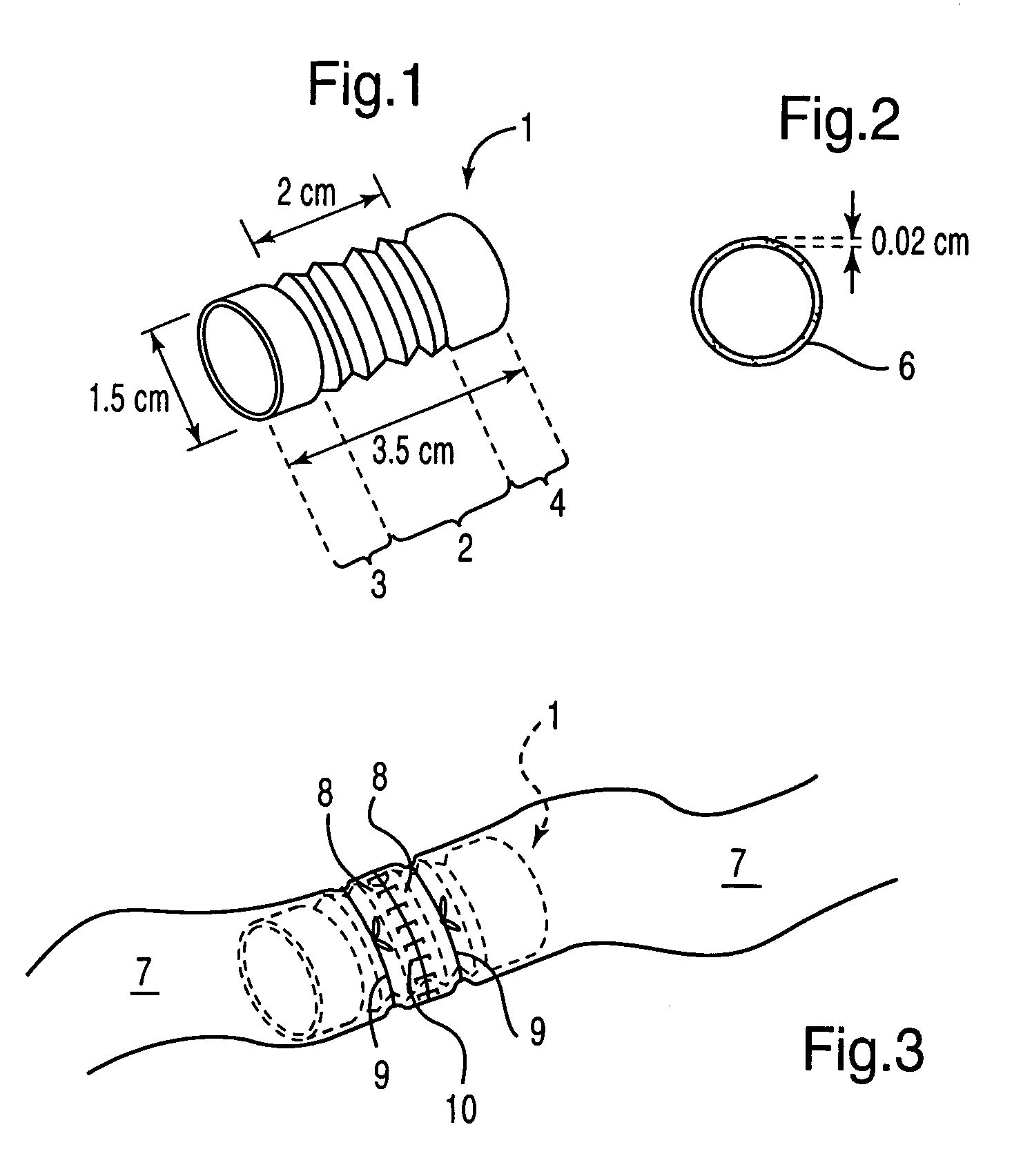
[0017]Referring to the drawings, wherein like reference numerals indicate like elements throughout the several figures, there is depicted a biocompatible and dissolvable stent 1 of the present invention for use in the anastomosis of a mammalian luminal viscus 7. A particularly applicable use of the present invention is for anastomosis of the bowel.

[0018]A stent in accordance with the present invention is comprised of a biocompatible material which safely dissolves in the body after a short period of time. The material must have sufficient strength and lasting power to allow the surgeon enough time to complete the anastomosis, but preferably of a material that will also completely and safely dissolve into the body after a short period of time thereafter. Furthermore, the material must also be soft enough to allow easy penetration by sutures or surgical staples. A particularly suitable biocompatible material for use in the invention is comprised of PGA.

[0019]As made apparent by FIGS. ...


PUM


Abstract

A temporary biocompatible stent and method for visceral anastomosis. The stent is provided with integral means for maintaining the structural stability of the stent while providing substantial flexibility. The method comprises fitting the luminal stumps of the viscus over either end of a stent of the invention, and joining the ends of each stump together. A short time after completion of the anastomosis, the stent dissolves and is absorbed safely into the body. The stent and method can be beneficially used in laparoscopic or more invasive traditional surgical procedures. The stent and method are particularly well suited for anastomosis of the bowel.

Description

CROSS REFERENCE TO RELATED APPLICATION[0001]This application is related to provisional application Ser. No. 60 / 132,692, filed May 4, 1999, which is incorporated herein by reference.BACKGROUND OF THE INVENTION[0002]1. Field of the Invention[0003]The procedure of choice to correct or relieve blocked or diseased luminal viscera of the body, such as the bowel, bile duct, ureter, and blood vessels in the medical field involves ligation and surgical anastomosis. Typically, a blocked or restricted luminal viscus, or a lesion or tumor on the viscus wall, is repaired whereby the abnormal portion is removed forming a break in the lumen. The break is then repaired by rejoining the two healthy luminal portions. Commonly, this was done by the surgeon with hand stitches or surgical staples. This procedure is performed on numerous tubular organs of the body, including the intestines and bowel, the bile duct, ureter, fallopian tubes, and blood vessels. These previous methods proved time consuming a...

Claims


Application Information

Patent Timeline
Patent Type & Authority Patents(United States)
IPC IPC(8): A61F2/06A61B17/03A61B17/08A61B17/11A61B17/115
CPCA61B17/11A61B17/1152A61B2017/1135
Inventor CHU, DAVID Z. J.
Owner CITY OF HOPE
Why Eureka
Try Eureka