
Advanced slow-magic angle spinning probe for magnetic resonance imaging and spectroscopy

a technology of magnetic resonance imaging and spectroscopy, applied in the field of new slow magic angle spinning probes, can solve the problems of affecting the qualitative and quantitative spectral analysis of static samples, hampering the quantitative, sometimes even qualitative, analysis of spectra, and often suffering from poor resolution

Inactive Publication Date: 2006-01-24
BATTELLE MEMORIAL INST
View PDF8 Cites 10 Cited by
  • Summary
  • Abstract
  • Description
  • Claims
  • Application Information

AI Technical Summary

Problems solved by technology

However, the 1H spectrum obtained from a static sample often suffers from poor resolution due to various line broadening mechanisms inherent within a biological system.
This hampers a quantitative, and sometimes even qualitative, analysis of the spectra.
This is especially true for 1H NMR spectroscopy where resulting spectra often contain severely overlapping spectral lines that can seriously hamper both the qualitative and quantitative spectral analysis.
Increasing the T2 weighting factors can sometimes enhance the spectral resolution, but can also result in serious signal losses and spectral distortions.
For example, if a lower spinning frequency is used, water SSBs can overlap with the metabolite lines rendering the interpretation of the spectra difficult.
A serious problem associated with conventional fast-MAS spectroscopy of fluid-filled objects (including live specimens) is the introduction of large centrifugal forces (Fc) that can be induced in a sample from the high spinning rates.
For example, spinning can destroy the tissue structure and even individual cells.
As an example, when F=2 kHz and r=1 cm, Fc can equal 1.6×10⁵ times the gravitational force G. Thus, standard MAS spectroscopic methods are not viable for the study of intact and / or larger biological objects, samples, or specimens and more particularly in vivo studies.
However, a slow, stable spin rate was not achieved in the modified commercial NMR probe over a sufficiently long time interval to be viable.
Further still, the commercial probe routinely overshot the upper frequency boundary limit of 100 Hz for safe spinning upon initial startup, which proved unacceptable for research involving live objects, including live animals.
Although these applications comprehensively describe the general procedures of the slow-MAS methodology, nowhere do they describe a slow-MAS probe suitable for applying the methodology to a fluid biological object.

Method used


Image

  • Advanced slow-magic angle spinning probe for magnetic resonance imaging and spectroscopy
  • Advanced slow-magic angle spinning probe for magnetic resonance imaging and spectroscopy
  • Advanced slow-magic angle spinning probe for magnetic resonance imaging and spectroscopy

Examples


example 1

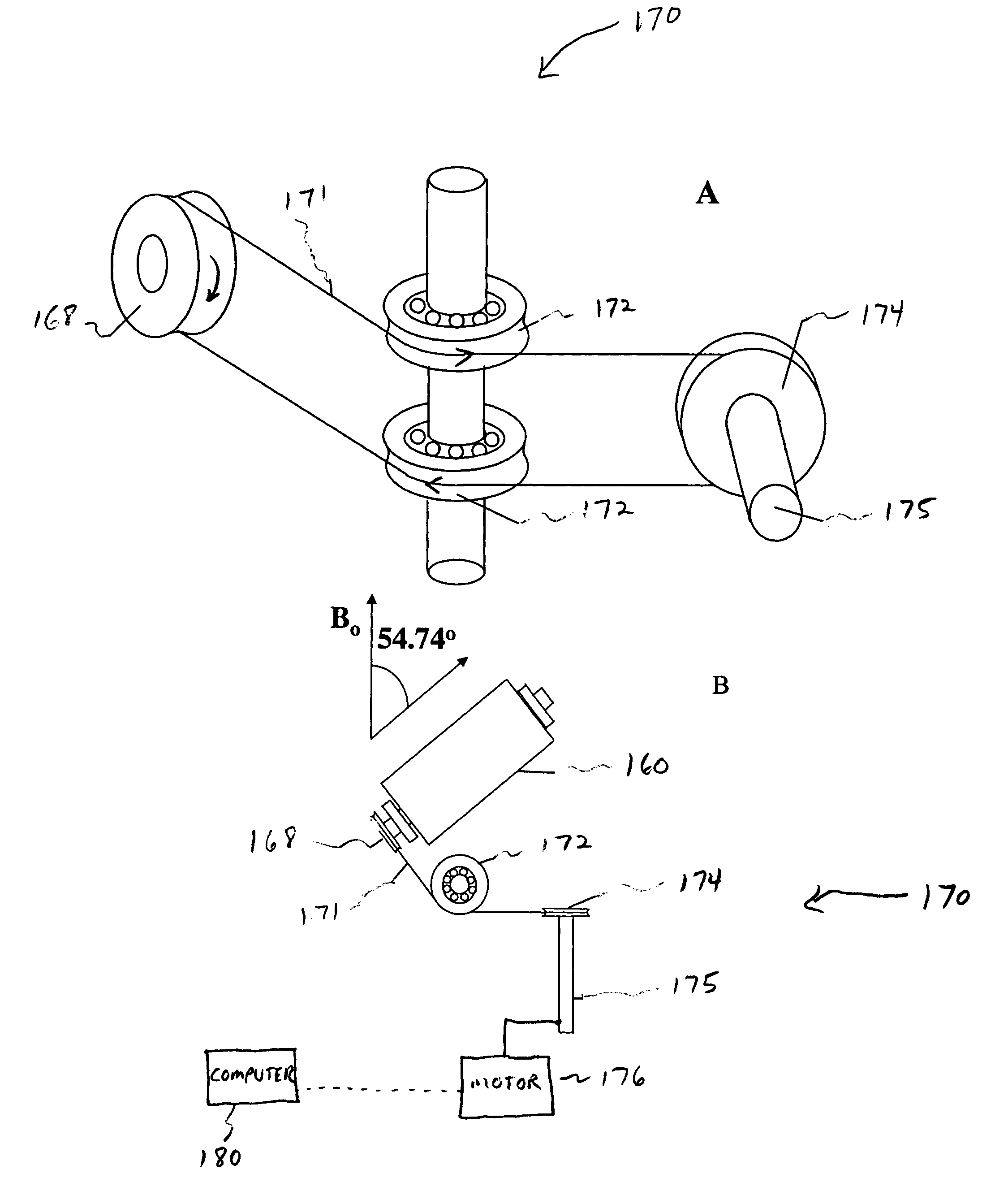
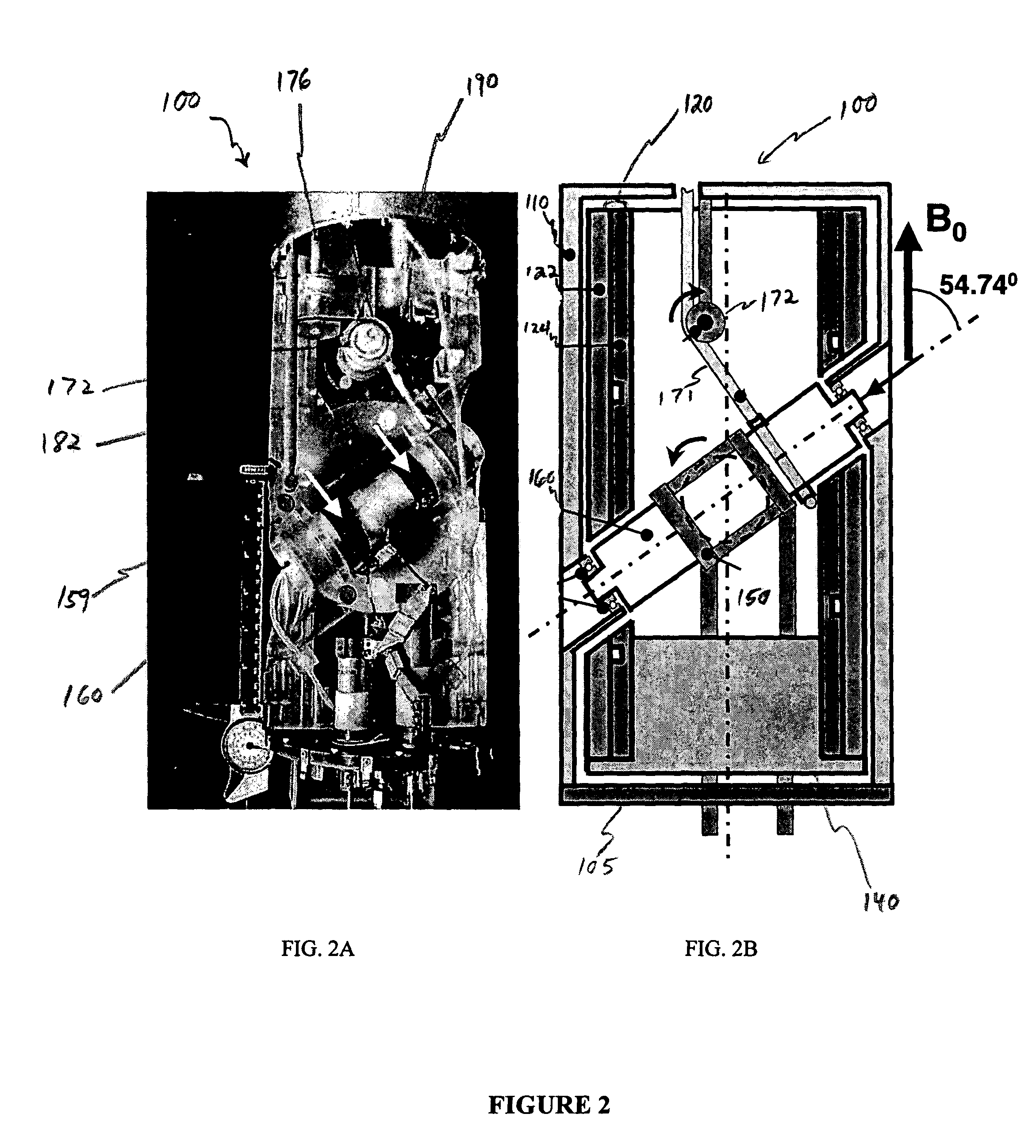
[0055]1H NMR experiments were performed using a horizontal wide-bore (30 cm) 2 Tesla Oxford magnet tuned to a proton Larmor frequency of 85 MHz, in combination with a Varian (Palo Alto, Calif.) Unity Plus console. In the present embodiment (FIG. 2A), the probe 100 had an O.D. of 120 mm, to fit in a standard gradient probe (not shown) inserted into the magnet. The rotor 160 comprised a custom-made (ARRK Product Development Group, Beaverton, Oreg.) epoxy photopolymer cylindrical mold 163 as described hereinabove and illustrated in FIG. 3. The mold 163 was cut into two halves to allow insertion of an animal specimen, in this case, a live, anesthetized female BALBc mouse. In the instant case, only the center part of the animal was probed and investigated. The outside diameter of the gradient probe was 220 mm and was shimmed using a shim probe (not shown) at room temperature. The rotor assembly 160 included a driving assembly 170 having a 1 / 12th HP motor (Baldor Series 18H AC Flux Vector...

example 2

[0058]FIG. 2B shows a picture of the probe 100 for a slow MAS experiment according to a second embodiment of the present invention. The probe 100 may be adapted and configured with incorporated gradient coils and with a longer rotor 160 to probe a whole body volume of a mouse positioned at the center of the magnet and the probe 100. For example, the outside diameter of the probe 100 without the gradients (FIG. 2A) is restricted to 120 mm, as it has to fit in a standard gradient probe with the same I.D. and an O.D. of 220 mm. By incorporating the gradients into the MAS probe, the standard gradient probe is no longer needed, which means that the diameter of the MAS-gradient probe can be increased to 220 mm. This is more than sufficient to perform research on all parts of a mouse, and probably a larger animal such as a rat as well. In fact, for a mouse MAS probe 100 the O.D. can be reduced to 150 mm, which makes it possible to mount this probe 100 in many high-field magnets (up to 12 T...

example 3

[0067]A freshly excised rat liver was analyzed using a 1 Hz PHORMAT experiment with a 7 T magnetic field. Results were compared with those previously obtained. In the present case (liver), the anisotropic line width of the 0.9 ppm peak was found to be 150 Hz, whereas in the mouse, a value of 60 Hz was observed in the 2 T field. As noted previously, the line broadening in biological objects is dominated by magnetic susceptibility and the anisotropic line width is approximately proportional to the external field. As a result, at 7 T, a line width of 210 Hz is predicted, which is larger than that found in the liver. The increased broadening in the mouse may be due to signals arising from tissues close to cavities like the lungs, where the susceptibility gradients are presumably larger than in the more homogeneous liver sample. The isotropic line width in the excised liver is 15 Hz, close to the 13 Hz width measured in the live mouse at 2 T. This is in agreement with the fact that the i...


PUM

PropertyMeasurementUnit
frequencyaaaaaaaaaa
frequencyaaaaaaaaaa
frequencyaaaaaaaaaa

Abstract

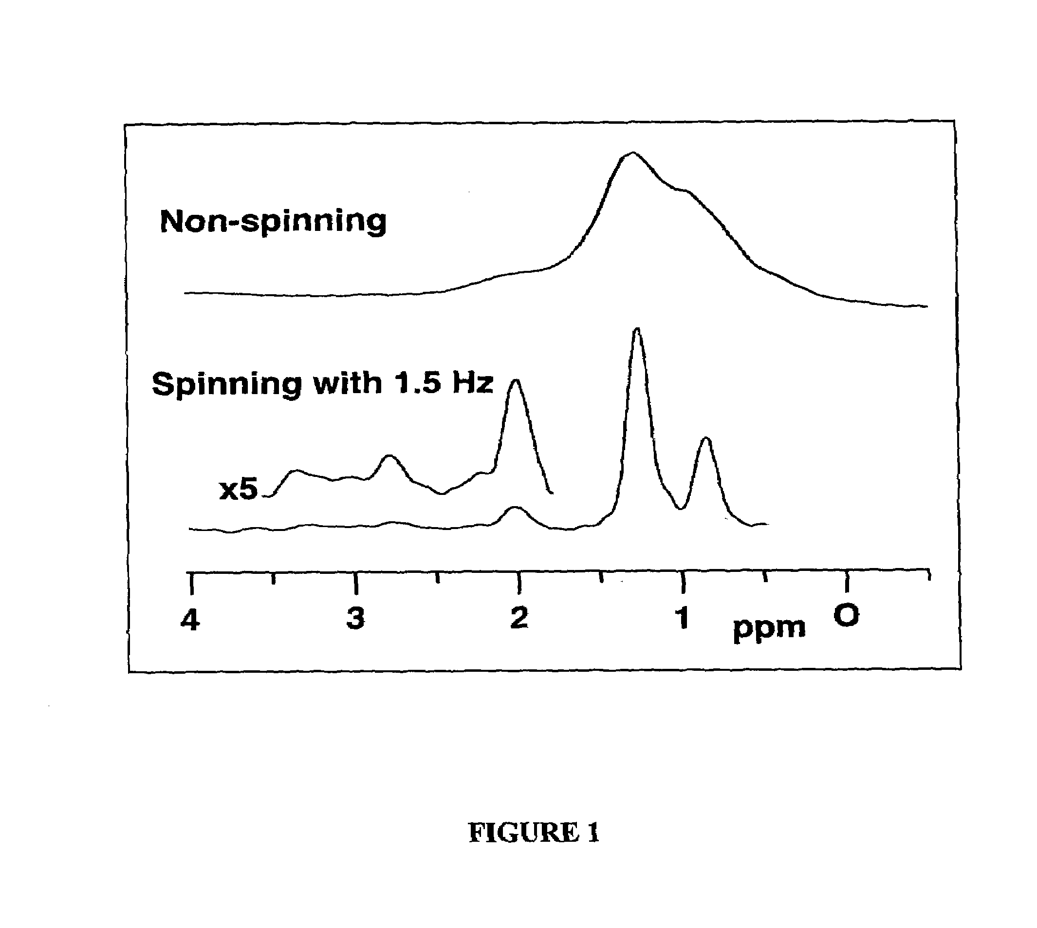
The present invention relates to a probe and processes useful for magnetic resonance imaging and spectroscopy instruments. More particularly, the invention relates to a MR probe and processes for obtaining resolution enhancements of fluid objects, including live specimens, using an ultra-slow (magic angle) spinning (MAS) of the specimen combined with a modified phase-corrected magic angle turning (PHORMAT) pulse sequence. Proton NMR spectra were measured of the torso and the top part of the belly of a female BALBc mouse in a 2T field, while spinning the animal at a speed of 1.5 Hz. Results show that even in this relatively low field with PHORMAT, an isotropic spectrum is obtained with line widths that are a factor 4.6 smaller than those obtained in a stationary mouse. Resolution of 1H NMR metabolite spectra are thus significantly enhanced. Results indicate that PHORMAT has the potential to significantly increase the utility of 1H NMR spectroscopy for in vivo biochemical, biomedical and / or medical applications involving large-sized biological objects such as mice, rats and even humans within a hospital setting. For small-sized objects, including biological objects, such as excised tissues, organs, live bacterial cells, and biofilms, use of PASS at a spinning rate of 30 Hz and above is preferred.

Description

[0001]The invention was made with Government support under Contract DE-AC0676RLO 1830, awarded by the U.S. Department of Energy. The Government has certain rights in the invention.BACKGROUND OF THE INVENTION[0002](1) Field of the Invention[0003]The present invention relates to a device for magnetic resonance (MR) imaging and spectroscopy of fluid-based objects, generally. More particularly, the present invention relates to a new slow “magic angle” spinning (slow-MAS) probe useful for Magnetic Resonance (MR) imaging and spectroscopy of biological objects, including live animals.[0004](2). Description of the Technical Art[0005]In vitro and in vivo 1H NMR spectroscopy are widely used for investigating biochemical processes in cells, tissues, animals, and humans (see, for example, I. R. Young, ed., methods in Biomedical magnetic resonance imaging and spectroscopy). However, the 1H spectrum obtained from a static sample often suffers from poor resolution due to various line broadening me...

Claims


Application Information

Patent Timeline
Patent Type & Authority Patents(United States)
IPC IPC(8): G01V3/00G01N24/08G01R33/30G01R33/46G01R33/465
CPCG01R33/307G01N24/08G01R33/465G01R33/4633B33Y80/00
Inventor WIND, ROBERT A.HU, JIAN ZHIMINARD, KEVIN R.ROMMEREIM, DONALD N.
Owner BATTELLE MEMORIAL INST
Learn More